The MTHFD1L Antibody (CAB7969) is a high-quality antibody developed for reliable detection and analysis of target proteins. This antibody, generated in rabbits, exhibits high specificity and reactivity towards the human MTHFD1L protein, making it ideal for use in Western blot experiments.MTHFD1L is crucial for the synthesis of purines and pyrimidines, essential building blocks for DNA and RNA production. Dysregulation of MTHFD1L has been linked to various diseases, including cancer, cardiovascular disorders, and neural tube defects.
This antibody is validated for use in WB, IHC-P, ELISA applications and has demonstrated reactivity against Human, Mouse, Rat samples.
Product Name:
MTHFD1L Antibody
SKU:
CAB7969
Size:
20μL, 100μL
Reactivity:
Human, Mouse, Rat
Conjugate:
Unconjugated
Immunogen:
Recombinant protein (or fragment).This information is considered to be commercially sensitive.
Recommended starting concentration is 1 μg/mL. Please optimize the concentration based on your specific assay requirements.
Synonyms:
FTHFSDC1, MTC1THFS, dJ292B18.2, MTHFD1L
Positive Sample:
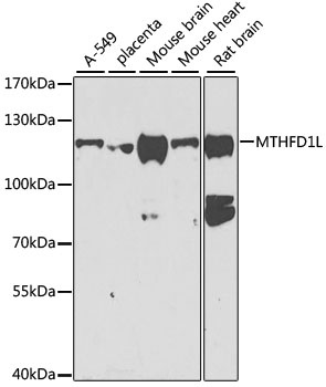
A-549, placenta, Mouse brain, Mouse heart, Rat brain
Cellular Localization:
Mitochondrion.
Calculated MW:
106kDa
Observed MW:
106kDa
The protein encoded by this gene is involved in the synthesis of tetrahydrofolate (THF) in the mitochondrion. THF is important in the de novo synthesis of purines and thymidylate and in the regeneration of methionine from homocysteine. Several transcript variants encoding different isoforms have been found for this gene.
Purification Method
Affinity purification
Gene ID
25902
RRID
AB_2770466
Buffer Information
Store at -20℃. Avoid freeze / thaw cycles. Buffer: PBS containing 50% glycerol, preserved with proclin300 or sodium azide, pH 7.3.
Western blot analysis of various lysates using MTHFD1L Rabbit pAb (CAB7969) at 1:1000 dilution. Secondary antibody: HRP-conjugated Goat anti-Rabbit IgG (H+L) (CABS014) at 1:10000 dilution. Lysates/proteins: 25μg per lane. Blocking buffer: 3% nonfat dry milk in TBST. Detection: ECL Basic Kit (AbGn00020). Exposure time: 90s.
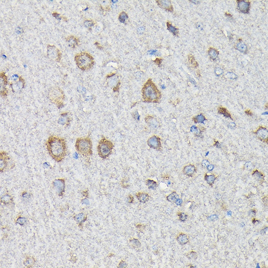
Immunohistochemistry analysis of paraffin-embedded Rat brain using MTHFD1L Rabbit pAb (CAB7969) at dilution of 1:200 (40x lens). High pressure antigen retrieval performed with 0.01M Citrate buffer (pH 6.0) prior to IHC staining.
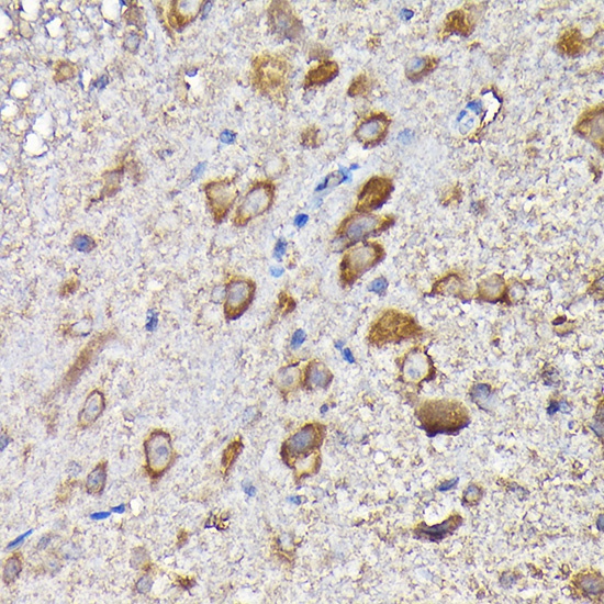
Immunohistochemistry analysis of paraffin-embedded Mouse spinal cord using MTHFD1L Rabbit pAb (CAB7969) at dilution of 1:200 (40x lens). High pressure antigen retrieval performed with 0.01M Citrate buffer (pH 6.0) prior to IHC staining.