The MTX2 Antibody (CAB7958) is a high-quality antibody developed for reliable detection and analysis of target proteins. This antibody, generated in rabbits, demonstrates high specificity and sensitivity for Metaxin-2 in human samples, making it a reliable choice for Western blot applications.Metaxin-2, also known as Mtx2, is a crucial component of the mitochondrial protein import machinery, facilitating the transport of proteins into the mitochondria for proper function. Research on Metaxin-2 is essential for understanding mitochondrial dynamics, metabolism, and cellular homeostasis.
This antibody is validated for use in WB, IF/ICC, ELISA applications and has demonstrated reactivity against Human, Mouse, Rat samples.
Product Name:
MTX2 Antibody
SKU:
CAB7958
Size:
20μL, 100μL
Reactivity:
Human, Mouse, Rat
Conjugate:
Unconjugated
Immunogen:
Recombinant protein (or fragment).This information is considered to be commercially sensitive.
Recommended starting concentration is 1 μg/mL. Please optimize the concentration based on your specific assay requirements.
Synonyms:
MDPS, metaxin-2, MTX2
Positive Sample:
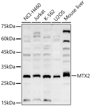
NCI-H460, Jurkat, K-562, U2OS, Mouse liver
Cellular Localization:
Mitochondrion, Mitochondrion Outer Membrane.
Calculated MW:
30kDa
Observed MW:
29kDa
The protein encoded by this gene is highly similar to the metaxin 2 protein from mouse, which has been shown to interact with the mitochondrial membrane protein metaxin 1. Because of this similarity, it is thought that the encoded protein is peripherally associated with the cytosolic face of the outer mitochondrial membrane, and that it is involved in the import of proteins into the mitochondrion. Alternative splicing results in multiple transcript variants. A related pseudogene has been identified on chromosome 7.
Purification Method
Affinity purification
Gene ID
10651
RRID
AB_2770482
Buffer Information
Store at -20℃. Avoid freeze / thaw cycles. Buffer: PBS containing 50% glycerol, preserved with proclin300 or sodium azide, pH 7.3.
Western blot analysis of various lysates, using MTX2 Rabbit pAb (CAB7958) at 1:2000 dilution. Secondary antibody: HRP-conjugated Goat anti-Rabbit IgG (H+L) (CABS014) at 1:10000 dilution. Lysates/proteins: 25μg per lane. Blocking buffer: 3% nonfat dry milk in TBST. Detection: ECL Basic Kit (AbGn00020). Exposure time: 180s.
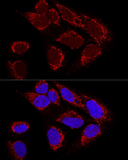
Confocal immunofluorescence analysis of U2OS cells using MTX2 Rabbit pAb (CAB7958) at dilution of 1:100. Blue: DAPI for nuclear staining.