The MYBBP1A Antibody (CAB4429) is a high-quality antibody developed for reliable detection and analysis of target proteins. This antibody, produced in rabbits, exhibits high specificity and sensitivity for detecting Mybbp1a in human samples, making it suitable for applications such as Western blotting.Mybbp1a is known to interact with transcription factors and chromatin-modifying enzymes, playing a crucial role in the regulation of gene expression.
This antibody is validated for use in WB, IHC-P, IF/ICC, ELISA applications and has demonstrated reactivity against Human, Mouse, Rat samples.
Product Name:
MYBBP1A Antibody
SKU:
CAB4429
Size:
20μL, 100μL
Reactivity:
Human, Mouse, Rat
Conjugate:
Unconjugated
Immunogen:
Recombinant protein (or fragment).This information is considered to be commercially sensitive.
Recommended starting concentration is 1 μg/mL. Please optimize the concentration based on your specific assay requirements.
Synonyms:
P160, PAP2, Pol5, MYBBP1A
Positive Sample:
HeLa, SW620, MCF7, Mouse liver, Rat liver
Cellular Localization:
Cytoplasm, Nucleus, Nucleolus.
Calculated MW:
149kDa
Observed MW:
149kDa
This gene encodes a nucleolar transcriptional regulator that was first identified by its ability to bind specifically to the Myb proto-oncogene protein. The encoded protein is thought to play a role in many cellular processes including response to nucleolar stress, tumor suppression and synthesis of ribosomal DNA. Alternate splicing results in multiple transcript variants.
Purification Method
Affinity purification
Gene ID
10514
RRID
AB_2765671
Buffer Information
Store at -20℃. Avoid freeze / thaw cycles. Buffer: PBS containing 50% glycerol, preserved with proclin300 or sodium azide, pH 7.3.
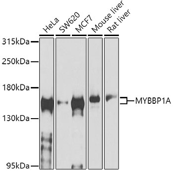
Western blot analysis of various lysates using MYBBP1A Rabbit pAb (CAB4429) at 1:1000 dilution. Secondary antibody: HRP-conjugated Goat anti-Rabbit IgG (H+L) (CABS014) at 1:10000 dilution. Lysates/proteins: 25μg per lane. Blocking buffer: 3% nonfat dry milk in TBST. Detection: ECL Basic Kit (AbGn00020). Exposure time: 60s.
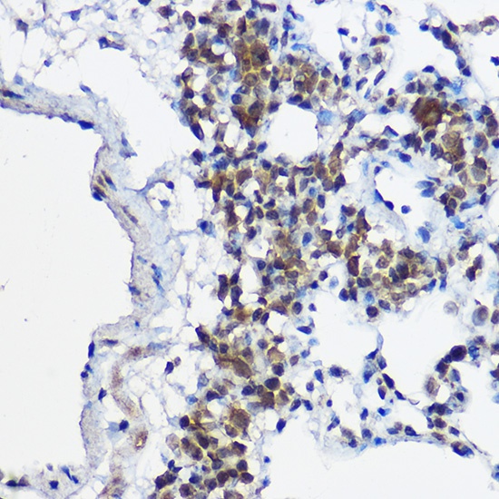
Immunohistochemistry analysis of paraffin-embedded Rat lung using MYBBP1A Rabbit pAb (CAB4429) at dilution of 1:100 (40x lens). High pressure antigen retrieval performed with 0.01M Citrate buffer (pH 6.0) prior to IHC staining.
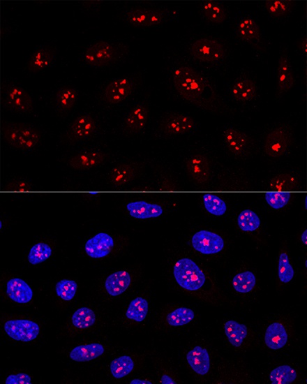
Confocal immunofluorescence analysis of U2OS cells using MYBBP1A Rabbit pAb (CAB4429) at dilution of 1:200. Blue: DAPI for nuclear staining.