The MYO1B Monoclonal Antibody (CAB21113) is a high-quality antibody developed for reliable detection and analysis of target proteins. Enables ATP binding activity; actin filament binding activity; and microfilament motor activity. Involved in actin filament organization and post-Golgi vesicle-mediated transport. Located in several cellular components, including actin filament; endosome; and perinuclear region of cytoplasm. Colocalizes with trans-Golgi network membrane.
This antibody is validated for use in WB, ELISA applications and has demonstrated reactivity against Human, Mouse, Rat samples.
Product Name:
MYO1B Monoclonal Antibody
SKU:
CAB21113
Size:
100μL, 20μL
Reactivity:
Human, Mouse, Rat
Clone Number:
ARC2990
Conjugate:
Unconjugated
Immunogen:
Recombinant protein (or fragment).This information is considered to be commercially sensitive.
Tested Applications:
WBELISA
Recommended Dilution:
WB
1:500 - 1:1000
ELISA
Recommended starting concentration is 1 μg/mL. Please optimize the concentration based on your specific assay requirements.
Synonyms:
MMIa, myr1, MYH-1c, MMI-alpha, MYO1B
Positive Sample:
A-431, Mouse lung, Rat lung
Cellular Localization:
Actin Cytoskeleton, Cytoplasm, Early Endosome, Extracellular Exosome, Filopodium, Perinuclear Region Of Cytoplasm, Plasma Membrane.
Calculated MW:
132kDa
Observed MW:
132kDa
Enables ATP binding activity; actin filament binding activity; and microfilament motor activity. Involved in actin filament organization and post-Golgi vesicle-mediated transport. Located in several cellular components, including actin filament; endosome; and perinuclear region of cytoplasm. Colocalizes with trans-Golgi network membrane.
Purification Method
Affinity purification
Gene ID
4430
Buffer Information
Store at -20℃. Avoid freeze / thaw cycles. Buffer: PBS containing 50% glycerol and 0.05% BSA, preserved with proclin300 or sodium azide, pH 7.3.
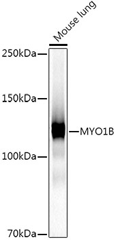
Western blot analysis of lysates from Mouse lung, using MYO1B Rabbit mAb (CAB21113) at1:1000 dilution. Secondary antibody: HRP-conjugated Goat anti-Rabbit IgG (H+L) (AS014) at 1:10000 dilution. Lysates/proteins: 25μg per lane. Blocking buffer: 3% nonfat dry milk in TBST. Detection: ECL Basic Kit (AbGn00020). Exposure time: 30s.
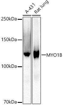
Western blot analysis of various lysates using MYO1B Rabbit mAb (CAB21113) at1:1000 dilution. Secondary antibody: HRP-conjugated Goat anti-Rabbit IgG (H+L) (AS014) at 1:10000 dilution. Lysates/proteins: 25μg per lane. Blocking buffer: 3% nonfat dry milk in TBST. Detection: ECL Basic Kit (AbGn00020). Exposure time: 180s.
 at1:1000 dilution. Secondary antibody: HRP Goat Anti-Rabbit IgG (H+L) at 1:10000 dilution. Lysates/proteins: 25μg per lane. Blocking buffer: 3% nonfat dry milk in TBST.")
 at1:1000 dilution. Secondary antibody: HRP Goat Anti-Rabbit IgG (H+L) at 1:10000 dilution. Lysates/proteins: 25μg per lane. Blocking buffer: 3% nonfat dry milk in TBST.")

")