The MYO1C Antibody (CAB6936) is a high-quality antibody developed for reliable detection and analysis of target proteins. This antibody, generated in rabbits, is highly sensitive and reactive with human samples, making it an ideal tool for studies in cell biology and cancer research.MYO1C, a member of the myosin superfamily of actin-based molecular motors, is involved in the movement and positioning of cellular membranes and organelles. Its dysregulation has been implicated in cancer progression and metastasis, making it a potential target for therapeutic interventions.
This antibody is validated for use in WB, IHC-P, IP, ELISA applications and has demonstrated reactivity against Human, Mouse, Rat samples.
Product Name:
MYO1C Antibody
SKU:
CAB6936
Size:
20μL, 100μL
Reactivity:
Human, Mouse, Rat
Conjugate:
Unconjugated
Immunogen:
Recombinant protein (or fragment).This information is considered to be commercially sensitive.
This gene encodes a member of the unconventional myosin protein family, which are actin-based molecular motors. The protein is found in the cytoplasm, and one isoform with a unique N-terminus is also found in the nucleus. The nuclear isoform associates with RNA polymerase I and II and functions in transcription initiation. The mouse ortholog of this protein also functions in intracellular vesicle transport to the plasma membrane. Multiple transcript variants encoding different isoforms have been found for this gene. The related gene myosin IE has been referred to as myosin IC in the literature, but it is a distinct locus on chromosome 19.
Purification Method
Affinity purification
Gene ID
4641
RRID
AB_2767494
Buffer Information
Store at -20℃. Avoid freeze / thaw cycles. Buffer: PBS containing 50% glycerol, preserved with proclin300 or sodium azide, pH 7.3.
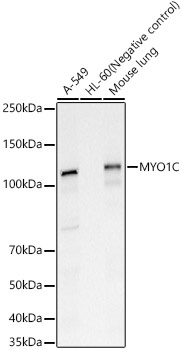
Western blot analysis of various lysates, using MYO1C Rabbit pAb (CAB6936) at 1:1500 dilution. Secondary antibody: HRP-conjugated Goat anti-Rabbit IgG (H+L) (CABS014) at 1:10000 dilution. Lysates/proteins: 25μg per lane. Blocking buffer: 3% nonfat dry milk in TBST. Detection: ECL Basic Kit (AbGn00020). Exposure time: 30s.
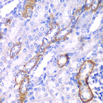
Immunohistochemistry analysis of paraffin-embedded Rat kidney using MYO1C Rabbit pAb (CAB6936) at dilution of 1:100 (40x lens). Microwave antigen retrieval performed with 0.01M PBS Buffer (pH 7.2) prior to IHC staining.
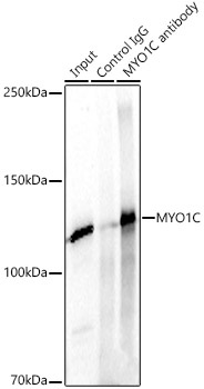
Immunoprecipitation analysis of 300ug extracts of A549 cells using 3ug MYO1C Rabbit pAb (CAB6936 1:100). Western blot was performed from the immunoprecipitate using MYO1C Rabbit pAb (CAB6936) at a dilition of 1:1000.