The MYH10 Antibody (CAB12029) is a high-quality antibody developed for reliable detection and analysis of target proteins. This gene encodes a member of the myosin superfamily. The protein represents a conventional non-muscle myosin; it should not be confused with the unconventional myosin-10 (MYO10). Myosins are actin-dependent motor proteins with diverse functions including regulation of cytokinesis, cell motility, and cell polarity. Mutations in this gene have been associated with May-Hegglin anomaly and developmental defects in brain and heart. Multiple transcript variants encoding different isoforms have been found for this gene.
This antibody is validated for use in WB, ELISA applications and has demonstrated reactivity against Human, Mouse, Rat samples.
Product Name:
MYH10 Antibody
SKU:
CAB12029
Size:
100μL, 20μL
Reactivity:
Human, Mouse, Rat
Conjugate:
Unconjugated
Immunogen:
Recombinant protein (or fragment).This information is considered to be commercially sensitive.
Tested Applications:
WBELISA
Recommended Dilution:
WB
1:500 - 1:2000
ELISA
Recommended starting concentration is 1 μg/mL. Please optimize the concentration based on your specific assay requirements.
Synonyms:
NMMHCB, NMMHC-IIB, MYH10
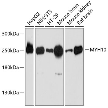
Positive Sample:
HepG2, NIH/3T3, HT-29, Mouse brain, Mouse kidney, Rat brain
Cellular Localization:
Cell Projection, Lamellipodium.
Calculated MW:
229kDa
Observed MW:
250kDa
This gene encodes a member of the myosin superfamily. The protein represents a conventional non-muscle myosin; it should not be confused with the unconventional myosin-10 (MYO10). Myosins are actin-dependent motor proteins with diverse functions including regulation of cytokinesis, cell motility, and cell polarity. Mutations in this gene have been associated with May-Hegglin anomaly and developmental defects in brain and heart. Multiple transcript variants encoding different isoforms have been found for this gene.
Purification Method
Affinity purification
Gene ID
4628
RRID
AB_2758939
Buffer Information
Store at -20℃. Avoid freeze / thaw cycles. Buffer: PBS with 0.01% thimerosal,50% glycerol,pH7.3.
Western blot analysis of various lysates using MYH10 Rabbit pAb (CAB12029) at 1:3000 dilution. Secondary antibody: HRP-conjugated Goat anti-Rabbit IgG (H+L) (AS014) at 1:10000 dilution. Lysates/proteins: 25μg per lane. Blocking buffer: 3% nonfat dry milk in TBST. Detection: ECL Basic Kit (AbGn00020). Exposure time: 30s.