The NAGA Antibody (CAB9942) is a high-quality antibody developed for reliable detection and analysis of target proteins. This antibody, produced in rabbits, exhibits high reactivity with human samples and has been validated for use in various applications, including Western blotting. By binding to the NAGA protein, this antibody enables precise detection and analysis in a range of cell types, making it ideal for studies in biochemistry and metabolism.NAGA, or alpha-N-acetylgalactosaminidase, plays a crucial role in lysosomal degradation pathways and is implicated in several metabolic disorders.
This antibody is validated for use in WB, IF/ICC, ELISA applications and has demonstrated reactivity against Human, Mouse, Rat samples.
Product Name:
NAGA Antibody
SKU:
CAB9942
Size:
20μL, 100μL
Reactivity:
Human, Mouse, Rat
Conjugate:
Unconjugated
Immunogen:
Recombinant protein (or fragment).This information is considered to be commercially sensitive.
Recommended starting concentration is 1 μg/mL. Please optimize the concentration based on your specific assay requirements.
Synonyms:
GALB, D22S674, NAGA
Positive Sample:
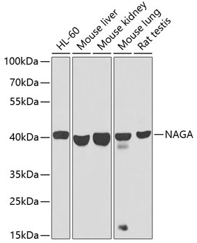
HL-60, Mouse liver, Mouse kidney, Mouse lung, Rat testis
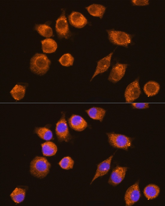
Cellular Localization:
Lysosome.
Calculated MW:
47kDa
Observed MW:
46kDa
NAGA encodes the lysosomal enzyme alpha-N-acetylgalactosaminidase, which cleaves alpha-N-acetylgalactosaminyl moieties from glycoconjugates. Mutations in NAGA have been identified as the cause of Schindler disease types I and II (type II also known as Kanzaki disease).
Purification Method
Affinity purification
Gene ID
4668
RRID
AB_2770515
Buffer Information
Store at -20℃. Avoid freeze / thaw cycles. Buffer: PBS containing 50% glycerol, preserved with proclin300 or sodium azide, pH 7.3.
Western blot analysis of various lysates using NAGA Rabbit pAb (CAB9942) at 1:1000 dilution. Secondary antibody: HRP-conjugated Goat anti-Rabbit IgG (H+L) (CABS014) at 1:10000 dilution. Lysates/proteins: 25μg per lane. Blocking buffer: 3% nonfat dry milk in TBST. Detection: ECL Basic Kit (AbGn00020). Exposure time: 30s.
Immunofluorescence analysis of L929 cells using NAGA Rabbit pAb (CAB9942) at dilution of 1:100 (40x lens). Secondary antibody: Cy3-conjugated Goat anti-Rabbit IgG (H+L) (CABS007) at 1:500 dilution. Blue: DAPI for nuclear staining.


 ELISA Kit (HUFI03337)")
 ELISA Kit (HUFI03087)")


