The [KO Validated] NBS1/NBN Antibody (CAB0783) is a high-quality antibody developed for reliable detection and analysis of target proteins. This antibody, raised in rabbits, is highly specific to human samples and has been validated for use in Western blot applications.The NBN protein, also known as nibrin, is part of the MRN complex involved in DNA double-strand break repair. Mutations in the NBN gene have been associated with a predisposition to certain types of cancer, making it a promising target for cancer research.
This antibody is validated for use in WB, IHC-P, ELISA applications and has demonstrated reactivity against Human, Mouse, Rat samples.
Product Name:
[KO Validated] NBS1/NBN Antibody
SKU:
CAB0783
Size:
20μL, 100μL
Reactivity:
Human, Mouse, Rat
Conjugate:
Unconjugated
Immunogen:
Recombinant protein (or fragment).This information is considered to be commercially sensitive.
Recommended starting concentration is 1 μg/mL. Please optimize the concentration based on your specific assay requirements.
Synonyms:
ATV, NBS, P95, NBS1, AT-V1, AT-V2, BN
Positive Sample:
293T, Mouse testis, Rat testis
Cellular Localization:
Chromosome, Nucleus, Pml Body, Telomere.
Calculated MW:
85kDa
Observed MW:
95kDa/70kDa
Mutations in this gene are associated with Nijmegen breakage syndrome, an autosomal recessive chromosomal instability syndrome characterized by microcephaly, growth retardation, immunodeficiency, and cancer predisposition. The encoded protein is a member of the MRE11/RAD50 double-strand break repair complex which consists of 5 proteins. This gene product is thought to be involved in DNA double-strand break repair and DNA damage-induced checkpoint activation.
Purification Method
Affinity purification
Gene ID
4683
RRID
AB_2757395
Buffer Information
Store at -20℃. Avoid freeze / thaw cycles. Buffer: PBS containing 50% glycerol, preserved with proclin300 or sodium azide, pH 7.3.
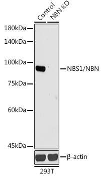
Western blot analysis of lysates from wild type (WT) and NBS1/NBS1/NBN knockout (KO) 293T cells, using [KO Validated] NBS1/NBN Rabbit pAb (CAB0783) at 1:1000 dilution. Secondary antibody: HRP-conjugated Goat anti-Rabbit IgG (H+L) (CABS014) at 1:10000 dilution. Lysates/proteins: 25μg per lane. Blocking buffer: 3% nonfat dry milk in TBST. Detection: ECL Basic Kit (AbGn00020). Exposure time: 90s.
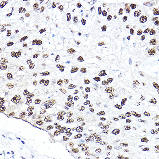
Immunohistochemistry analysis of paraffin-embedded Human lung cancer using [KO Validated] NBS1/NBS1/NBN Rabbit pAb (CAB0783) at dilution of 1:50 (40x lens). High pressure antigen retrieval performed with 0.01M Citrate buffer (pH 6.0) prior to IHC staining.