The NDUFB2 Antibody (CAB3978) is a high-quality antibody developed for reliable detection and analysis of target proteins. This antibody, generated in rabbits, exhibits high reactivity with human samples and has been validated for use in Western blot applications. By specifically binding to the NDUFB2 protein, this antibody enables accurate detection and analysis in a variety of cell types, making it an ideal choice for studies in mitochondrial biology, energy metabolism, and related research areas.NDUFB2 is essential for the function of complex I in the electron transport chain, which is responsible for generating the majority of cellular ATP.
This antibody is validated for use in WB, ELISA applications and has demonstrated reactivity against Human, Mouse, Rat samples.
Product Name:
NDUFB2 Antibody
SKU:
CAB3978
Size:
20μL, 100μL
Reactivity:
Human, Mouse, Rat
Conjugate:
Unconjugated
Immunogen:
Recombinant protein (or fragment).This information is considered to be commercially sensitive.
The protein encoded by this gene is a subunit of the multisubunit NADH:ubiquinone oxidoreductase (complex I). Mammalian complex I is composed of 45 different subunits. This protein has NADH dehydrogenase activity and oxidoreductase activity. It plays a important role in transfering electrons from NADH to the respiratory chain. The immediate electron acceptor for the enzyme is believed to be ubiquinone. Hydropathy analysis revealed that this subunit and 4 other subunits have an overall hydrophilic pattern, even though they are found within the hydrophobic protein (HP) fraction of complex I.
Purification Method
Affinity purification
Gene ID
4708
RRID
AB_2765425
Buffer Information
Store at -20℃. Avoid freeze / thaw cycles. Buffer: PBS with 0.01% thimerosal,50% glycerol,pH7.3.
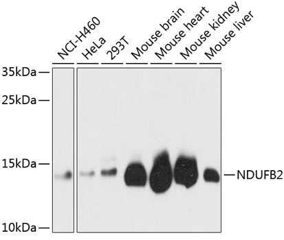
Western blot analysis of various lysates using NDUFB2 Rabbit pAb (CAB3978) at 1:3000 dilution. Secondary antibody: HRP-conjugated Goat anti-Rabbit IgG (H+L) (CABS014) at 1:10000 dilution. Lysates/proteins: 25μg per lane. Blocking buffer: 3% nonfat dry milk in TBST. Detection: ECL Basic Kit (AbGn00020). Exposure time: 30s.