The NeuN Monoclonal Antibody (CAB19086) is a high-quality antibody developed for reliable detection and analysis of target proteins. This rabbit monoclonal antibody is highly specific and sensitive for Neun in human samples, making it an ideal choice for Western blot applications. Neun is a neuron-specific RNA-binding protein that plays a critical role in neuronal development and function. It is commonly used as a marker for post-mitotic neurons in various research applications, including neuroscience, neurodegenerative diseases, and brain development studies.
This antibody is validated for use in WB, IHC-P, ELISA, IF-F, IF-P, mIHC applications and has demonstrated reactivity against Human, Mouse, Rat samples.
Product Name:
NeuN Monoclonal Antibody
SKU:
CAB19086
Size:
20μL, 100μL
Reactivity:
Human, Mouse, Rat
Clone Number:
ARC0202
Conjugate:
Unconjugated
Immunogen:
Synthetic peptide. This information is considered to be commercially sensitive.
Recommended starting concentration is 1 μg/mL. Please optimize the concentration based on your specific assay requirements.
Synonyms:
FOX3, NEUN, FOX-3, HRNBP3, NeuN
Positive Sample:
Mouse brain, Rat brain
Cellular Localization:
Cytoplasm, Nucleus.
Calculated MW:
34kDa
Observed MW:
46-55kDa
This gene encodes a member of the RNA-binding FOX protein family which is involved in the regulation of alternative splicing of pre-mRNA. The protein has an N-terminal proline-rich region, an RNA recognition motif (RRM) domain, and a C-terminal alanine-rich region. This gene produces the neuronal nuclei (NeuN) antigen that has been widely used as a marker for post-mitotic neurons. This gene has its highest expression in the central nervous system and plays a prominent role in neural tissue development and regulation of adult brain function. Mutations in this gene have been associated with numerous neurological disorders. Alternative splicing of this gene results in multiple transcript variants encoding distinct isoforms.
Purification Method
Affinity purification
Gene ID
146713
RRID
AB_2862578
Buffer Information
Store at -20℃. Avoid freeze / thaw cycles. Buffer: PBS with 0.09% sodium azide,0.05% BSA,50% glycerol,pH7.3.
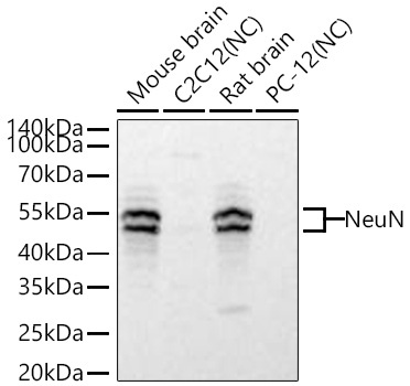
Western blot analysis of various lysates using NeuN Rabbit mAb (CAB19086) at 1:5000 dilution incubated overnight at 4℃. Secondary antibody: HRP-conjugated Goat anti-Rabbit IgG (H+L) (CABS014) at 1:10000 dilution. Lysates/proteins: 25 μg per lane. Blocking buffer: 3% nonfat dry milk in TBST. Detection: ECL Basic Kit (AbGn00020). Negative control (NC): C2C12,PC-12 Exposure time: 10s.
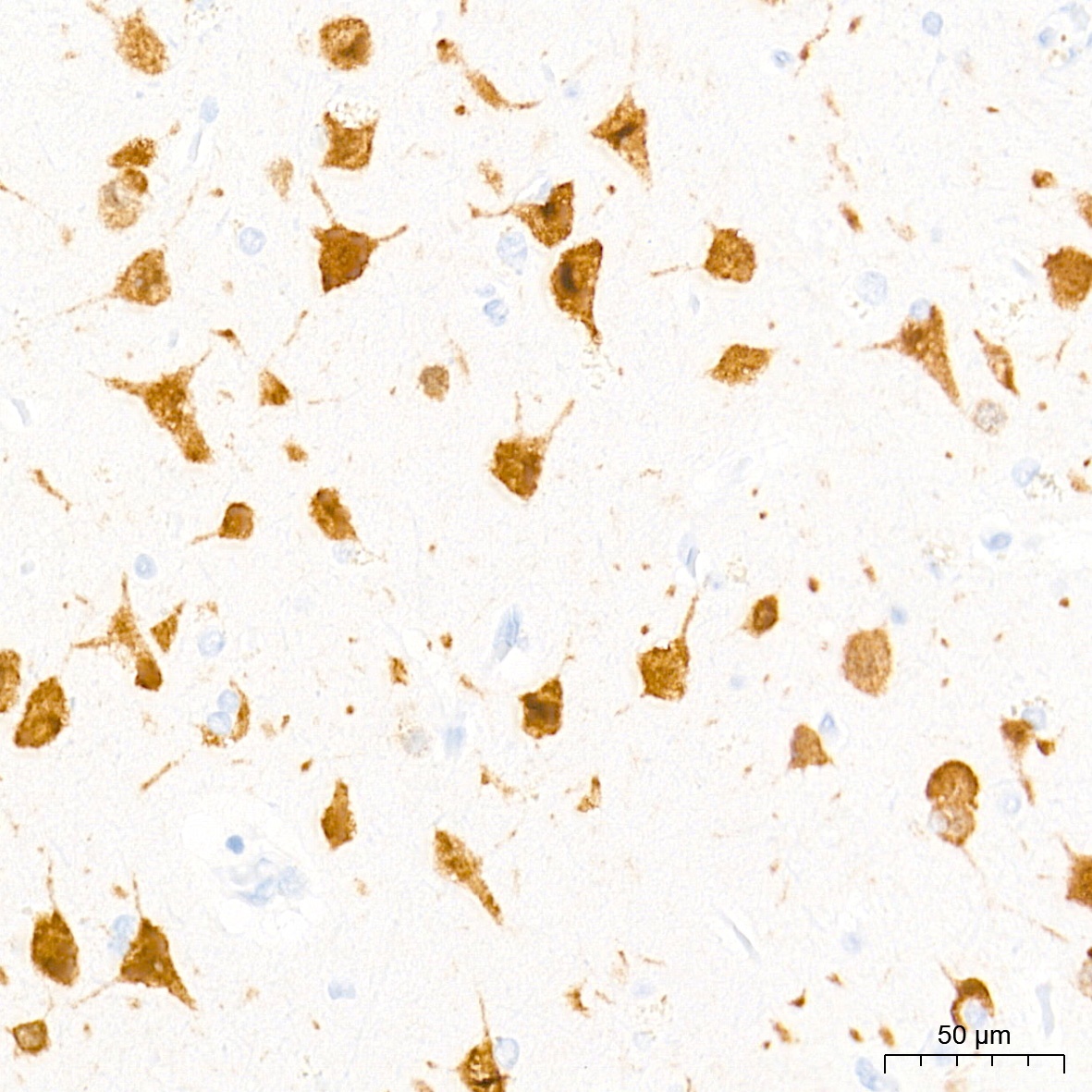
Immunohistochemistry analysis of paraffin-embedded Human brain tissue using NeuN Rabbit mAb (CAB19086) at a dilution of 1:2000 (40x lens). High pressure antigen retrieval performed with 0.01M Tris-EDTA Buffer (pH 9.0) prior to IHC staining.
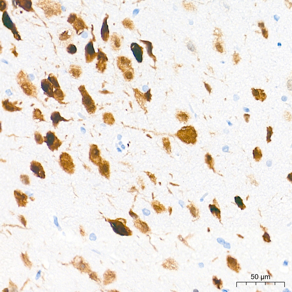
Immunohistochemistry analysis of paraffin-embedded Rat brain tissue using NeuN Rabbit mAb (CAB19086) at a dilution of 1:2000 (40x lens). High pressure antigen retrieval performed with 0.01M Tris-EDTA Buffer (pH 9.0) prior to IHC staining.
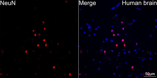
Confocal imaging of paraffin-embedded Human brain tissue using NeuN Rabbit mAb (CAB19086, dilution 1:200) followed by a further incubation with Cy3 Goat Anti-Rabbit IgG (H+L) (CABS007, dilution 1:500) (Red). DAPI was used for nuclear staining (Blue). High pressure antigen retrieval performed with 0.01M Citrate Buffer (pH 6.0) prior to IF staining. Objective: 40x.
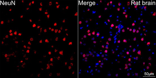
Confocal imaging of paraffin-embedded Rat brain tissue using NeuN Rabbit mAb (CAB19086, dilution 1:200) followed by a further incubation with Cy3 Goat Anti-Rabbit IgG (H+L) (CABS007, dilution 1:500) (Red). DAPI was used for nuclear staining (Blue). High pressure antigen retrieval performed with 0.01M Citrate Buffer (pH 6.0) prior to IF staining. Objective: 40x.
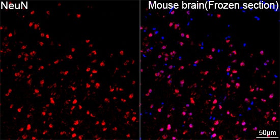
Confocal imaging of frozen sections Mouse brain(Frozen section) tissue using NeuN Rabbit mAb (CAB19086, dilution 1:200) followed by a further incubation with Cy3 Goat Anti-Rabbit IgG (H+L) (CABS007, dilution 1:500) (Red). DAPI was used for nuclear staining (Blue). Microwave antigen retrieval performed with 0.01M Citrate Buffer (pH 6.0) prior to IF staining. Objective: 40x.
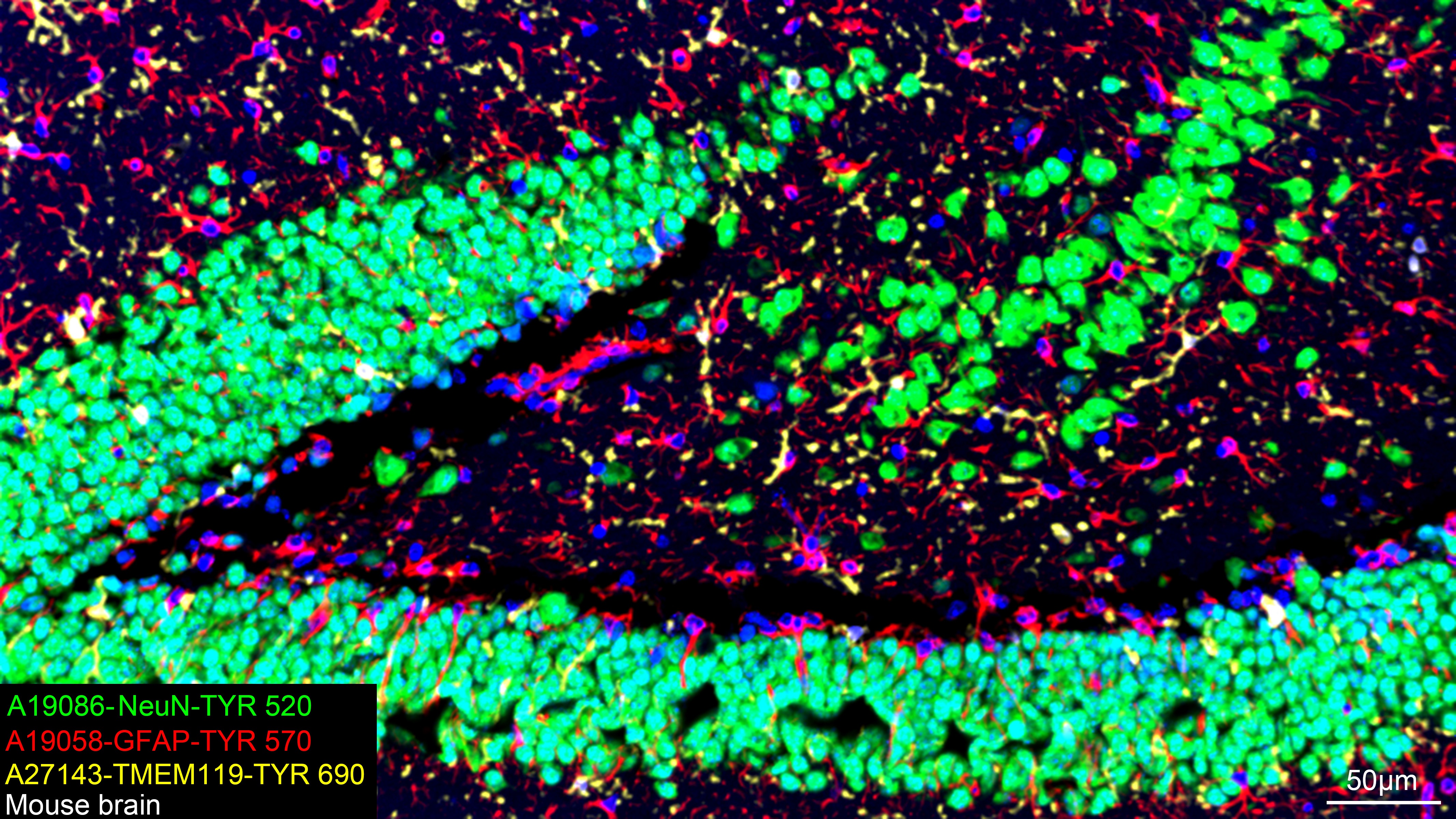
The multiplex IHC analysis on paraffin-embedded Mouse brain tissue using the following specific primary antibodies and tyramide signal amplification (TSA) reagents (RK05903) : NeuN Rabbit mAb (CAB19086, 1:2000) with TSA-TYR-520 (Green), GFAP Rabbit mAb (CAB19058, 1:500) with TSA-TYR-570 (Red), and TMEM119 Rabbit mAb (CAB27143, 1:600) with TSA-TYR-690 (Yellow). DAPI (Blue) was used for nuclear staining. Prior to multiplex IHC staining, high-pressure antigen retrieval was performed using 0.01M citrate buffer at pH 6.0. The analysis was completed using a 20x objective lens.