The Neuropilin-1 (NRP1) Monoclonal Antibody (CAB22213) is a high-quality antibody developed for reliable detection and analysis of target proteins. This monoclonal antibody, developed through rigorous testing and validation, specifically targets the NRP1 protein and is optimized for use in various research applications, such as immunofluorescence and flow cytometry.Neuropilin-1 is a critical player in tumor development, making this antibody a valuable tool for cancer research. Its expression on immune cells also suggests a role in immune regulation, opening up avenues for exploration in immunology and autoimmune disease studies.
This antibody is validated for use in WB, IF/ICC, ELISA applications and has demonstrated reactivity against Human samples.
Product Name:
Neuropilin-1 (NRP1) Monoclonal Antibody
SKU:
CAB22213
Size:
20μL, 100μL
Reactivity:
Human
Clone Number:
ARC55415
Conjugate:
Unconjugated
Immunogen:
Recombinant protein (or fragment).This information is considered to be commercially sensitive.
Cell Membrane, Secreted, Single-Pass Type I Membrane Protein.
Calculated MW:
68kDa/71kDa/103kDa
Observed MW:
120-140kDa
This gene encodes one of two neuropilins, which contain specific protein domains which allow them to participate in several different types of signaling pathways that control cell migration. Neuropilins contain a large N-terminal extracellular domain, made up of complement-binding, coagulation factor V/VIII, and meprin domains. These proteins also contains a short membrane-spanning domain and a small cytoplasmic domain. Neuropilins bind many ligands and various types of co-receptors; they affect cell survival, migration, and attraction. Some of the ligands and co-receptors bound by neuropilins are vascular endothelial growth factor (VEGF) and semaphorin family members. This protein has also been determined to act as a co-receptor for SARS-CoV-2 (which causes COVID-19) to infect host cells.
Purification Method
Affinity purification
Gene ID
8829
Buffer Information
Store at -20℃. Avoid freeze / thaw cycles. Buffer: PBS containing 50% glycerol and 0.05% BSA, preserved with proclin300 or sodium azide, pH 7.3.
Western blot analysis of various lysates using Neuropilin-1 (NRP1) Rabbit mAb (CAB22213) at 1:5000 dilution incubated at room temperature for 1.5 hours. Secondary antibody: HRP-conjugated Goat anti-Rabbit IgG (H+L) (CABS014) at 1:10000 dilution. Lysates/proteins: 25 μg per lane. Blocking buffer: 3% nonfat dry milk in TBST. Detection: ECL Basic Kit (AbGn00020). Negative control (NC): BxPC-3. Exposure time: 60 s.
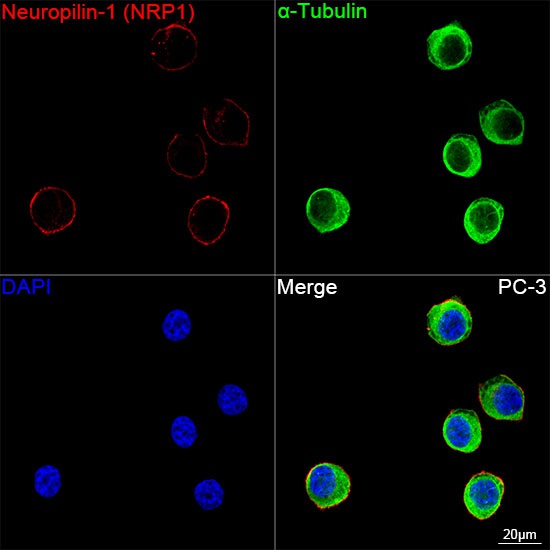
Confocal imaging of PC-3 cells using Neuropilin-1 (NRP1) Rabbit mAb (CAB22213, dilution 1:3000) followed by a further incubation with Cy3-conjugated Goat anti-Rabbit IgG (H+L) (CABS007, dilution 1:500) (Red). The cells were counterstained with α-Tubulin Mouse mAb (AC012, dilution 1:400) followed by incubation with ABflo® 488-conjugated Goat Anti-Mouse IgG (H+L) Ab (CABS076, dilution 1:500) (Green). DAPI was used for nuclear staining (Blue). Objective: 100x.
 antibody (CAB22213) at1:2000 dilution. Secondary antibody: HRP Goat Anti-Rabbit IgG (H+L) at 1:200000 dilution. Lysates/proteins: 25μg per lane. Blocking buffer: 3% nonfat dry milk in TBST.")

 antibody (CAB22213) at1:2000 dilution. Secondary antibody: HRP Goat Anti-Rabbit IgG (H+L) at 1:200000 dilution. Lysates/proteins: 25μg per lane. Blocking buffer: 3% nonfat dry milk in TBST.")


 ELISA Kit")
 ELISA Kit (AEES00551)")