The NFATC2 Antibody (CAB3107) is a high-quality antibody developed for reliable detection and analysis of target proteins. This antibody is sourced from rabbits and demonstrates high reactivity with human samples, making it suitable for use in techniques such as Western blotting.NFATC2, also known as Nuclear Factor of Activated T-cells 2, plays a crucial role in the regulation of genes involved in the immune system, making it a significant target for research in immunology and cancer biology. By binding specifically to NFATC2 protein, this antibody enables precise detection and analysis in various cell types, providing insights into the function and regulation of this transcription factor.
This antibody is validated for use in WB, IHC-P, IF/ICC, ELISA, IF-P applications and has demonstrated reactivity against Human, Mouse, Rat samples.
Product Name:
NFATC2 Antibody
SKU:
CAB3107
Size:
20μL, 100μL
Reactivity:
Human, Mouse, Rat
Conjugate:
Unconjugated
Immunogen:
Recombinant protein (or fragment).This information is considered to be commercially sensitive.
Recommended starting concentration is 1 μg/mL. Please optimize the concentration based on your specific assay requirements.
Synonyms:
JCOSL, NFAT1, NFATP, NFATC2
Positive Sample:
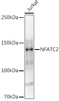
Jurkat
Cellular Localization:
Cytoplasm, Nucleus.
Calculated MW:
100kDa
Observed MW:
140kDa
This gene is a member of the nuclear factor of activated T cells (NFAT) family. The product of this gene is a DNA-binding protein with a REL-homology region (RHR) and an NFAT-homology region (NHR). This protein is present in the cytosol and only translocates to the nucleus upon T cell receptor (TCR) stimulation, where it becomes a member of the nuclear factors of activated T cells transcription complex. This complex plays a central role in inducing gene transcription during the immune response. Alternate transcriptional splice variants encoding different isoforms have been characterized.
Purification Method
Affinity purification
Gene ID
4773
RRID
AB_2764905
Buffer Information
Store at -20℃. Avoid freeze / thaw cycles. Buffer: PBS containing 50% glycerol, preserved with proclin300 or sodium azide, pH 7.3.
Western blot analysis of lysates from Jurkat cells, using (CAB3107) at 1:1000 dilution. Secondary antibody: HRP-conjugated Goat anti-Rabbit IgG (H+L) (CABS014) at 1:10000 dilution. Lysates/proteins: 25μg per lane. Blocking buffer: 3% nonfat dry milk in TBST. Detection: ECL Basic Kit (AbGn00020). Exposure time: 30s.
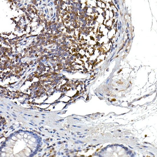
Immunohistochemistry analysis of paraffin-embedded Human colon using NFATC2 Rabbit pAb (CAB3107) at dilution of 1:100 (40x lens). High pressure antigen retrieval performed with 0.01M Citrate buffer (pH 6.0) prior to IHC staining.
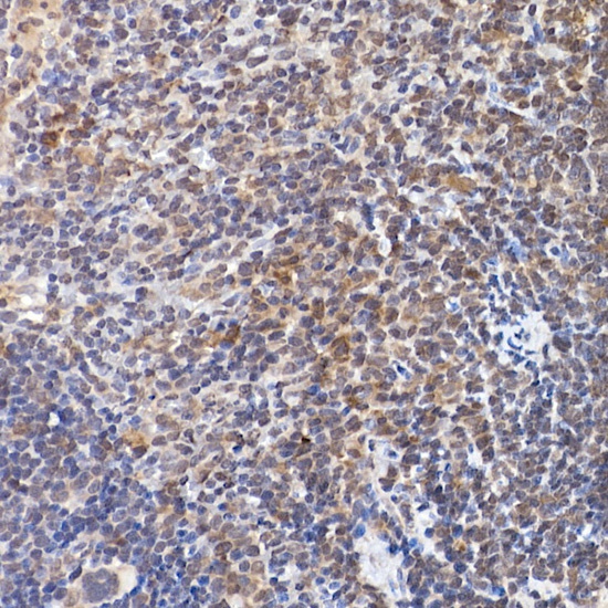
Immunohistochemistry analysis of paraffin-embedded Mouse spleen using NFATC2 Rabbit pAb (CAB3107) at dilution of 1:100 (40x lens). High pressure antigen retrieval performed with 0.01M Citrate buffer (pH 6.0) prior to IHC staining.
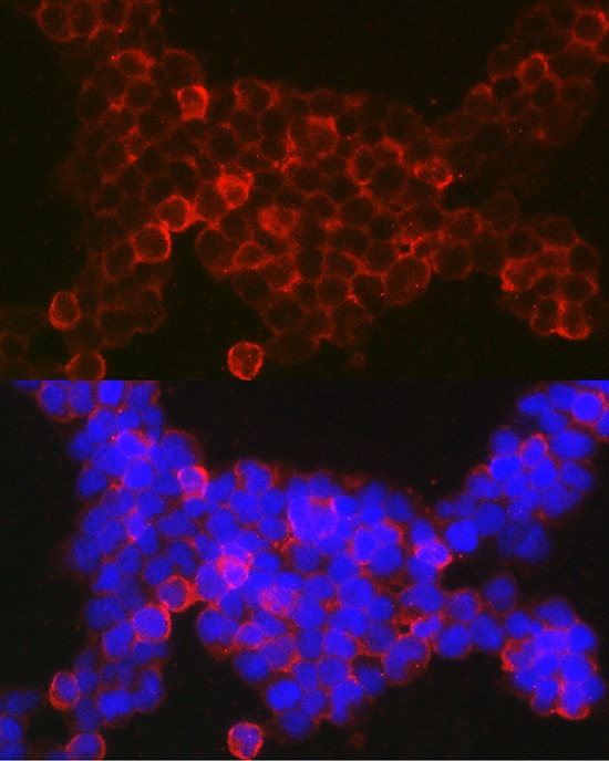
Immunofluorescence analysis of Jurkat cells using NFATC2 Rabbit pAb (CAB3107) at dilution of 1:100 (40x lens). Secondary antibody: Cy3-conjugated Goat anti-Rabbit IgG (H+L) (CABS007) at 1:500 dilution. Blue: DAPI for nuclear staining.