The NFS1 Antibody (CAB6668) is a high-quality antibody developed for reliable detection and analysis of target proteins. This antibody, developed in rabbits, specifically targets NFS1 in human samples and is validated for use in Western blot applications. By binding to the NFS1 protein, this antibody allows for the accurate detection and analysis of NFS1 levels in a variety of cell types, making it a useful resource for studies in biochemistry and molecular biology.NFS1 is essential for the proper functioning of several important cellular processes, including DNA repair, regulation of gene expression, and oxidative stress response.
This antibody is validated for use in WB, IHC-P, IF/ICC, ELISA applications and has demonstrated reactivity against Human, Mouse, Rat samples.
Product Name:
NFS1 Antibody
SKU:
CAB6668
Size:
20μL, 100μL
Reactivity:
Human, Mouse, Rat
Conjugate:
Unconjugated
Immunogen:
Recombinant protein (or fragment).This information is considered to be commercially sensitive.
Recommended starting concentration is 1 μg/mL. Please optimize the concentration based on your specific assay requirements.
Synonyms:
IscS, NIFS, COXPD52, HUSSY-08, NFS1
Positive Sample:
SH-SY5Y, HepG2, BxPC-3, Mouse brain, Mouse liver, Mouse heart, Rat brain, Rat liver, Rat heart
Cellular Localization:
Cytoplasm, Mitochondrion, Nucleus.
Calculated MW:
50kDa
Observed MW:
47kDa
Iron-sulfur clusters are required for the function of many cellular enzymes. The proteins encoded by this gene supply inorganic sulfur to these clusters by removing the sulfur from cysteine, creating alanine in the process. This gene uses alternate in-frame translation initiation sites to generate mitochondrial forms and cytoplasmic/nuclear forms. Selection of the alternative initiation sites is determined by the cytosolic pH. The encoded proteins belong to the class-V family of pyridoxal phosphate-dependent aminotransferases. Alternatively spliced transcript variants have been described.
Purification Method
Affinity purification
Gene ID
9054
RRID
AB_2767254
Buffer Information
Store at -20℃. Avoid freeze / thaw cycles. Buffer: PBS with 0.09% Sodium azide,50% glycerol,pH7.3.
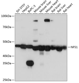
Western blot analysis of various lysates using NFS1 Rabbit pAb (CAB6668) at 1:1000 dilution. Secondary antibody: HRP-conjugated Goat anti-Rabbit IgG (H+L) (CABS014) at 1:10000 dilution. Lysates/proteins: 25μg per lane. Blocking buffer: 3% nonfat dry milk in TBST. Detection: ECL Basic Kit (AbGn00020). Exposure time: 3s.
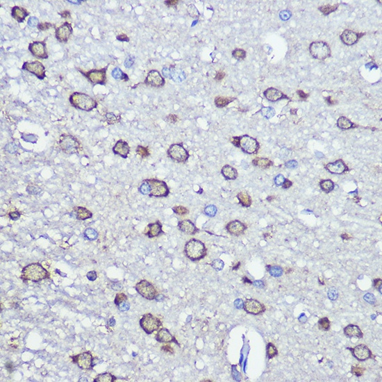
Immunohistochemistry analysis of paraffin-embedded Rat brain using NFS1 Rabbit pAb (CAB6668) at dilution of 1:100 (40x lens). Microwave antigen retrieval performed with 0.01M PBS Buffer (pH 7.2) prior to IHC staining.
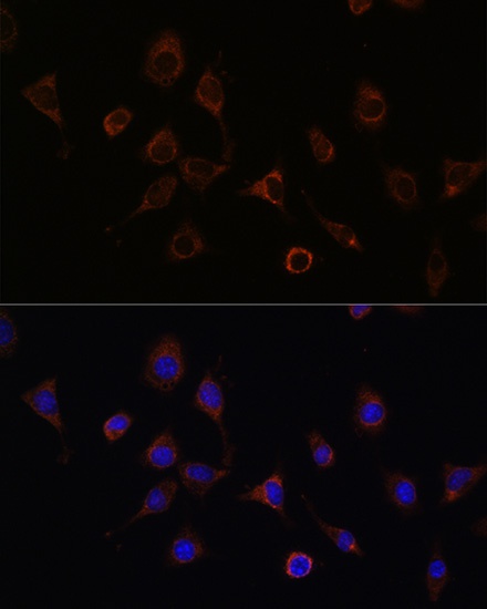
Immunofluorescence analysis of L929 cells using NFS1 Rabbit pAb (CAB6668) at dilution of 1:100 (40x lens). Secondary antibody: Cy3-conjugated Goat anti-Rabbit IgG (H+L) (CABS007) at 1:500 dilution. Blue: DAPI for nuclear staining.
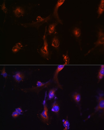
Immunofluorescence analysis of U-251 MG cells using NFS1 Rabbit pAb (CAB6668) at dilution of 1:100 (40x lens). Secondary antibody: Cy3-conjugated Goat anti-Rabbit IgG (H+L) (CABS007) at 1:500 dilution. Blue: DAPI for nuclear staining.