The NINJ1 Antibody (CAB16406) is a high-quality antibody developed for reliable detection and analysis of target proteins. This rabbit-derived antibody is highly specific to human samples and has been validated for use in Western blot applications. By binding to the NINJ1 protein, this antibody enables the detection and analysis of NINJ1 in a variety of cell types, making it ideal for investigations in immunology and cancer research.NINJ1, also known as Neuronal Growth Regulator 1, plays a crucial role in promoting cell survival and neurite outgrowth, making it a potential target for therapeutic interventions in neurodegenerative disorders and nerve regeneration.
This antibody is validated for use in WB, ELISA applications and has demonstrated reactivity against Human, Mouse, Rat samples.
Product Name:
NINJ1 Antibody
SKU:
CAB16406
Size:
20μL, 100μL
Reactivity:
Human, Mouse, Rat
Conjugate:
Unconjugated
Immunogen:
Synthetic peptide. This information is considered to be commercially sensitive.
Recommended starting concentration is 1 μg/mL. Please optimize the concentration based on your specific assay requirements.
Synonyms:
NIN1, NINJURIN, NINJ1
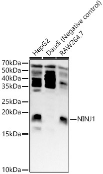
Positive Sample:
HepG2, Daudi, RAW264.7
Cellular Localization:
Membrane, Multi-Pass Membrane Protein.
Calculated MW:
16kDa
Observed MW:
18kDa
The ninjurin protein is upregulated after nerve injury both in dorsal root ganglion neurons and in Schwann cells (Araki and Milbrandt, 1996 [PubMed 8780658]). It demonstrates properties of a homophilic adhesion molecule and promotes neurite outgrowth from primary cultured dorsal root ganglion neurons.
Purification Method
Affinity purification
Gene ID
4814
RRID
AB_2770588
Buffer Information
Store at -20℃. Avoid freeze / thaw cycles. Buffer: PBS with 0.09% Sodium azide,50% glycerol,pH7.3.
Western blot analysis of various lysates, using NINJ1 Rabbit pAb (CAB16406) at 1:400 dilution. Secondary antibody: HRP-conjugated Goat anti-Rabbit IgG (H+L) (CABS014) at 1:10000 dilution. Lysates/proteins: 25μg per lane. Blocking buffer: 3% nonfat dry milk in TBST. Detection: ECL Basic Kit (AbGn00020). Exposure time: 60s.