The NOS2 Antibody (CAB14031) is a high-quality antibody developed for reliable detection and analysis of target proteins. This antibody, generated in rabbits, has high specificity for human samples and has been validated for use in Western blot applications.NOS2 plays a crucial role in the regulation of immune responses, particularly in the context of inflammation and host defense against pathogens. By targeting NOS2 with this antibody, researchers can gain valuable insights into the function and regulation of this enzyme in various cell types.
This antibody is validated for use in WB, IHC-P, IF/ICC, ELISA applications and has demonstrated reactivity against Human, Mouse, Rat samples.
Product Name:
NOS2 Antibody
SKU:
CAB14031
Size:
20μL, 100μL
Reactivity:
Human, Mouse, Rat
Conjugate:
Unconjugated
Immunogen:
Recombinant protein (or fragment).This information is considered to be commercially sensitive.
Recommended starting concentration is 1 μg/mL. Please optimize the concentration based on your specific assay requirements.
Synonyms:
NOS, INOS, NOS2A, HEP-NOS, iNOS
Positive Sample:
RAW 264.7 treated with LPS
Cellular Localization:
Cortical Cytoskeleton, Cytoplasm, Cytosol, Nucleoplasm, Nucleus, Perinuclear Region Of Cytoplasm, Peroxisomal Matrix, Peroxisome, Plasma Membrane.
Calculated MW:
131kDa
Observed MW:
130kDa
Nitric oxide is a reactive free radical which acts as a biologic mediator in several processes, including neurotransmission and antimicrobial and antitumoral activities. This gene encodes a nitric oxide synthase which is expressed in liver and is inducible by a combination of lipopolysaccharide and certain cytokines. Three related pseudogenes are located within the Smith-Magenis syndrome region on chromosome 17.
Purification Method
Affinity purification
Gene ID
4843
RRID
AB_2760886
Buffer Information
Store at -20℃. Avoid freeze / thaw cycles. Buffer: PBS with 0.09% Sodium azide,50% glycerol,pH7.3.
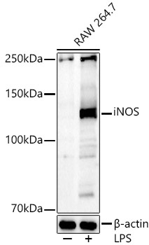
Western blot analysis of lysates from RAW264.7 cells, using iNOS Rabbit pAb (CAB14031) at 1:700 dilution. Raw264. 7 cells were treated with LPS (1 μg/ml) at 37℃ for 8 hours. Secondary antibody: HRP-conjugated Goat anti-Rabbit IgG (H+L) (CABS014) at 1:10000 dilution. Lysates/proteins: 25μg per lane. Blocking buffer: 3% nonfat dry milk in TBST. Detection: ECL Basic Kit (AbGn00020). Exposure time: 60s.
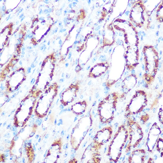
Immunohistochemistry analysis of paraffin-embedded Mouse kidney using iNOS Rabbit pAb (CAB14031) at dilution of 1:100 (40x lens). Microwave antigen retrieval performed with 0.01M Tris/EDTA Buffer (pH 9.0) prior to IHC staining.


 ELISA Kit (HUFI03337)")
 ELISA Kit (HUFI03087)")


