The Notch1 Antibody (CAB16673) is a high-quality antibody developed for reliable detection and analysis of target proteins. This gene encodes a member of the NOTCH family of proteins. Members of this Type I transmembrane protein family share structural characteristics including an extracellular domain consisting of multiple epidermal growth factor-like (EGF) repeats, and an intracellular domain consisting of multiple different domain types. Notch signaling is an evolutionarily conserved intercellular signaling pathway that regulates interactions between physically adjacent cells through binding of Notch family receptors to their cognate ligands. The encoded preproprotein is proteolytically processed in the trans-Golgi network to generate two polypeptide chains that heterodimerize to form the mature cell-surface receptor. This receptor plays a role in the development of numerous cell and tissue types. Mutations in this gene are associated with aortic valve disease, Adams-Oliver syndrome, T-cell acute lymphoblastic leukemia, chronic lymphocytic leukemia, and head and neck squamous cell carcinoma. RRID AB_2770619 Gene ID 4851 Swiss Prot Synonym hN1; AOS5; TAN1; AOVD1; Notch1
This antibody is validated for use in WB, IHC-P, IF/ICC, ELISA applications and has demonstrated reactivity against Human, Mouse, Rat samples.
Product Name:
Notch1 Antibody
SKU:
CAB16673
Size:
100μL, 20μL
Reactivity:
Human, Mouse, Rat
Clone Number:
-
Conjugate:
Unconjugated
Immunogen:
Recombinant protein (or fragment).This information is considered to be commercially sensitive.
Tested Applications:
WBIHC-PIF/ICCELISA
Recommended Dilution:
WB
1:500 - 1:2000
IHC-P
1:50 - 1:200
IF
/
ICC
1:50 - 1:200
ELISA
Recommended starting concentration is 1 μg/mL. Please optimize the concentration based on your specific assay requirements.
Synonyms:
hN1, AOS5, TAN1, AOVD1, Notch1
Positive Sample:
U-251MG
Cellular Localization:
Cell Membrane, Nucleus, Single-Pass Type I Membrane Protein.
Calculated MW:
273kDa
Observed MW:
120kDa
This gene encodes a member of the NOTCH family of proteins. Members of this Type I transmembrane protein family share structural characteristics including an extracellular domain consisting of multiple epidermal growth factor-like (EGF) repeats, and an intracellular domain consisting of multiple different domain types. Notch signaling is an evolutionarily conserved intercellular signaling pathway that regulates interactions between physically adjacent cells through binding of Notch family receptors to their cognate ligands. The encoded preproprotein is proteolytically processed in the trans-Golgi network to generate two polypeptide chains that heterodimerize to form the mature cell-surface receptor. This receptor plays a role in the development of numerous cell and tissue types. Mutations in this gene are associated with aortic valve disease, Adams-Oliver syndrome, T-cell acute lymphoblastic leukemia, chronic lymphocytic leukemia, and head and neck squamous cell carcinoma. RRID AB_2770619 Gene ID 4851 Swiss Prot Synonym hN1; AOS5; TAN1; AOVD1; Notch1
Purification Method:
Affinity purification
Gene ID:
4851
RRID:
AB_2770619
Buffer Information:
Store at -20℃. Avoid freeze / thaw cycles. Buffer: PBS with 0.09% sodium azide,50% glycerol,pH7.3.
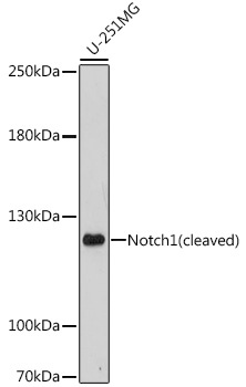
Western blot analysis of lysates from U-251MG cells, using Notch1 Rabbit pAb (CAB16673) at 1:1000 dilution. Secondary antibody: HRP-conjugated Goat anti-Rabbit IgG (H+L) (AS014) at 1:10000 dilution. Lysates/proteins: 25μg per lane. Blocking buffer: 3% nonfat dry milk in TBST. Detection: ECL Enhanced Kit (AbGn00021). Exposure time: 300s.
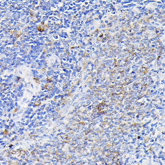
Immunohistochemistry analysis of paraffin-embedded Rat spleen using Notch1 Rabbit pAb (CAB16673) at dilution of 1:100 (40x lens). High pressure antigen retrieval performed with 0.01M Citrate buffer (pH 6.0) prior to IHC staining.
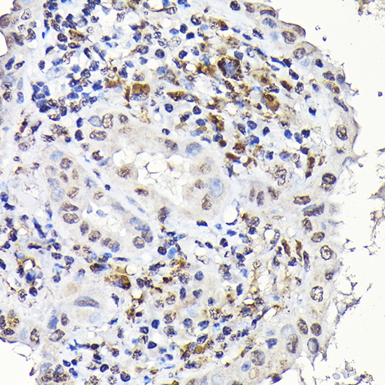
Immunohistochemistry analysis of paraffin-embedded Human thyroid cancer using Notch1 Rabbit pAb (CAB16673) at dilution of 1:100 (40x lens). High pressure antigen retrieval performed with 0.01M Citrate buffer (pH 6.0) prior to IHC staining.
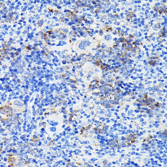
Immunohistochemistry analysis of paraffin-embedded Mouse spleen using Notch1 Rabbit pAb (CAB16673) at dilution of 1:100 (40x lens). High pressure antigen retrieval performed with 0.01M Citrate buffer (pH 6.0) prior to IHC staining.
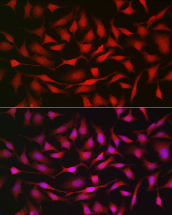
Immunofluorescence analysis of HeLa cells using Notch1 Rabbit pAb (CAB16673) at dilution of 1:200 (40x lens). Secondary antibody: Cy3-conjugated Goat anti-Rabbit IgG (H+L) (AS007) at 1:500 dilution. Blue: DAPI for nuclear staining.
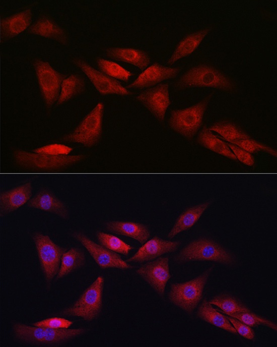
Immunofluorescence analysis of NIH/3T3 cells using Notch1 Rabbit pAb (CAB16673) at dilution of 1:200 (40x lens). Secondary antibody: Cy3-conjugated Goat anti-Rabbit IgG (H+L) (AS007) at 1:500 dilution. Blue: DAPI for nuclear staining.
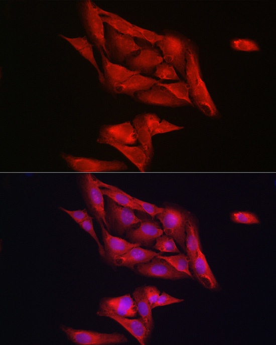
Immunofluorescence analysis of U2OS cells using Notch1 Rabbit pAb (CAB16673) at dilution of 1:200 (40x lens). Secondary antibody: Cy3-conjugated Goat anti-Rabbit IgG (H+L) (AS007) at 1:500 dilution. Blue: DAPI for nuclear staining.