The NQO1 Antibody (CAB1518) is a high-quality antibody developed for reliable detection and analysis of target proteins. This antibody, produced in rabbits, exhibits high specificity and sensitivity for human samples, making it an ideal choice for Western blot applications.NQO1, also known as DT-diaphorase or quinone reductase 1, plays a crucial role in protecting cells from oxidative stress and maintaining cellular redox balance. Its aberrant expression has been linked to various diseases, including cancer, neurodegenerative disorders, and cardiovascular diseases.
This antibody is validated for use in WB, IHC-P, IF/ICC, ELISA applications and has demonstrated reactivity against Human, Mouse, Rat samples.
Product Name:
NQO1 Antibody
SKU:
CAB1518
Size:
20μL, 100μL
Reactivity:
Human, Mouse, Rat
Conjugate:
Unconjugated
Immunogen:
Recombinant protein (or fragment).This information is considered to be commercially sensitive.
Recommended starting concentration is 1 μg/mL. Please optimize the concentration based on your specific assay requirements.
Synonyms:
DTD, QR1, DHQU, DIA4, NMOR1, NMORI, NQO1
Positive Sample:
HepG2, MCF7, NIH/3T3,Rat liver
Cellular Localization:
Cytoplasm.
Calculated MW:
31kDa
Observed MW:
31kDa
This gene is a member of the NAD(P)H dehydrogenase (quinone) family and encodes a cytoplasmic 2-electron reductase. This FAD-binding protein forms homodimers and reduces quinones to hydroquinones. This protein's enzymatic activity prevents the one electron reduction of quinones that results in the production of radical species. Mutations in this gene have been associated with tardive dyskinesia (TD), an increased risk of hematotoxicity after exposure to benzene, and susceptibility to various forms of cancer. Altered expression of this protein has been seen in many tumors and is also associated with Alzheimer's disease (AD). Alternate transcriptional splice variants, encoding different isoforms, have been characterized.
Purification Method
Affinity purification
Gene ID
1728
RRID
AB_2762070
Buffer Information
Store at -20℃. Avoid freeze / thaw cycles. Buffer: PBS containing 50% glycerol, preserved with proclin300 or sodium azide, pH 7.3.
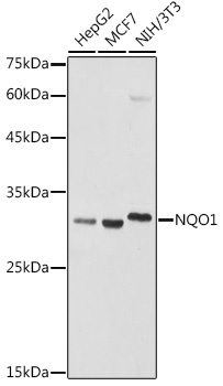
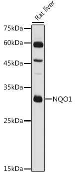
Western blot analysis of various lysates using NQO1 Rabbit pAb (CAB1518) at 1:1000 dilution. Secondary antibody: HRP-conjugated Goat anti-Rabbit IgG (H+L) (CABS014) at 1:10000 dilution. Lysates/proteins: 25μg per lane. Blocking buffer: 3% nonfat dry milk in TBST. Detection: ECL Basic Kit (AbGn00020). Exposure time: 1s.
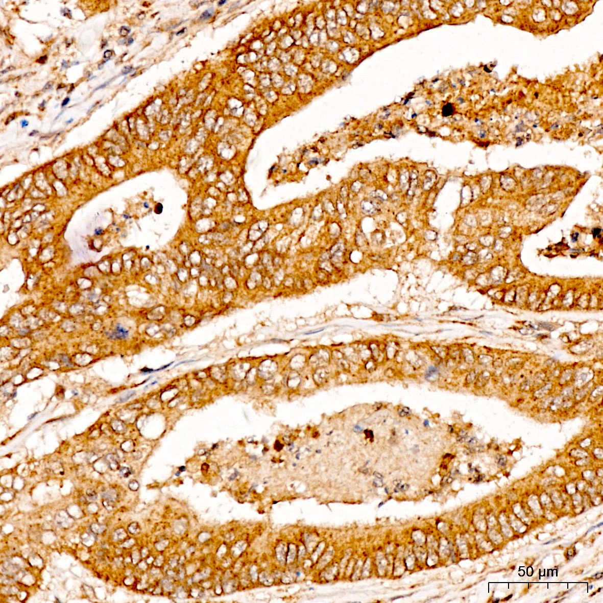
Immunohistochemistry analysis of paraffin-embedded Human colon carcinoma tissue using NQO1 Rabbit pAb (CAB1518) at a dilution of 1:100 (40x lens). High pressure antigen retrieval was performed with 0.01 M citrate buffer (pH 6.0) prior to IHC staining.
Immunohistochemistry analysis of paraffin-embedded Human colon carcinoma tissue using NQO1 Rabbit pAb (CAB1518) at a dilution of 1:100 (40x lens). High pressure antigen retrieval was performed with 0.01 M citrate buffer (pH 6.0) prior to IHC staining.
Immunohistochemistry analysis of paraffin-embedded Human liver tissue using NQO1 Rabbit pAb (CAB1518) at a dilution of 1:100 (40x lens). High pressure antigen retrieval was performed with 0.01 M citrate buffer (pH 6.0) prior to IHC staining.