The NQO1 Monoclonal Antibody (CAB19586) is a high-quality antibody developed for reliable detection and analysis of target proteins. This antibody, developed using rabbit monoclonal technology, exhibits high specificity and sensitivity towards human samples, making it a reliable choice for various applications including Western blotting.NQO1 is a versatile enzyme involved in the metabolism of various endogenous and exogenous compounds, playing a crucial role in cellular protection against oxidative stress and carcinogenesis. Dysregulation of NQO1 has been linked to numerous diseases, including cancer, neurodegenerative disorders, and metabolic syndromes, making it a promising target for therapeutic interventions and biomarker discovery.
This antibody is validated for use in WB, ELISA, IF-P applications and has demonstrated reactivity against Human, Mouse, Rat samples.
Product Name:
NQO1 Monoclonal Antibody
SKU:
CAB19586
Size:
20μL, 100μL
Reactivity:
Human, Mouse, Rat
Clone Number:
ARC56753
Conjugate:
Unconjugated
Immunogen:
Synthetic peptide. This information is considered to be commercially sensitive.
This gene is a member of the NAD(P)H dehydrogenase (quinone) family and encodes a cytoplasmic 2-electron reductase. This FAD-binding protein forms homodimers and reduces quinones to hydroquinones. This protein's enzymatic activity prevents the one electron reduction of quinones that results in the production of radical species. Mutations in this gene have been associated with tardive dyskinesia (TD), an increased risk of hematotoxicity after exposure to benzene, and susceptibility to various forms of cancer. Altered expression of this protein has been seen in many tumors and is also associated with Alzheimer's disease (AD). Alternate transcriptional splice variants, encoding different isoforms, have been characterized.
Purification Method
Affinity purification
Gene ID
1728
RRID
AB_2862684
Buffer Information
Store at -20℃. Avoid freeze / thaw cycles. Buffer: PBS containing 50% glycerol and 0.05% BSA, preserved with proclin300 or sodium azide, pH 7.3.
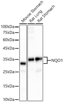
Western blot analysis of various lysates, using [KO Validated] NQO1 Rabbit mAb (CAB19586) at 1:20000 dilution. Secondary antibody: HRP-conjugated Goat anti-Rabbit IgG (H+L) (CABS014) at 1:10000 dilution. Lysates/proteins: 25μg per lane. Blocking buffer: 3% nonfat dry milk in TBST. Detection: ECL Basic Kit (AbGn00020). Exposure time: 10s.
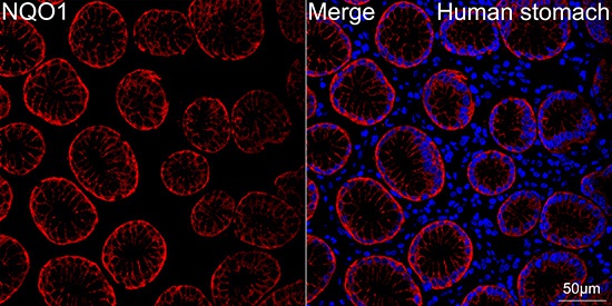
Confocal imaging of paraffin-embedded Human stomach tissue using [KO Validated] NQO1 Rabbit mAb (CAB19586, dilution 1:300) followed by a further incubation with Cy3 Goat Anti-Rabbit IgG (H+L) (CABS007, dilution 1:500) (Red). DAPI was used for nuclear staining (Blue). High pressure antigen retrieval performed with 0.01M Citrate Buffer (pH 6.0) prior to IF staining. Objective: 40x.
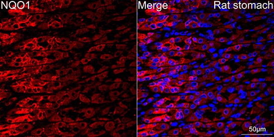
Confocal imaging of paraffin-embedded Rat stomach tissue using [KD Validated] NQO1 Rabbit mAb (CAB19586, dilution 1:300) followed by a further incubation with Cy3 Goat Anti-Rabbit IgG (H+L) (CABS007, dilution 1:500) (Red). DAPI was used for nuclear staining (Blue). High pressure antigen retrieval performed with 0.01M Citrate Buffer (pH 6.0) prior to IF staining. Objective: 40x.