The NRBF2 Antibody (CAB6462) is a high-quality antibody developed for reliable detection and analysis of target proteins. This antibody, generated in rabbits, is specifically designed for detecting NRBF2 in human samples through Western blot applications. Its high reactivity and specificity make it an ideal choice for investigating the role of NRBF2 in various cellular processes.NRBF2, also known as Nuclear Receptor Binding Factor 2, is a key player in the autophagy pathway, which is essential for maintaining cellular homeostasis and responding to stress.
This antibody is validated for use in WB, IHC-P, IF/ICC, ELISA applications and has demonstrated reactivity against Human, Mouse, Rat samples.
Product Name:
NRBF2 Antibody
SKU:
CAB6462
Size:
20μL, 100μL
Reactivity:
Human, Mouse, Rat
Conjugate:
Unconjugated
Immunogen:
Recombinant protein (or fragment).This information is considered to be commercially sensitive.
Involved in autophagy. Located in cytoplasm. Colocalizes with phosphatidylinositol 3-kinase complex, class III.
Purification Method
Affinity purification
Gene ID
29982
RRID
AB_2767064
Buffer Information
Store at -20℃. Avoid freeze / thaw cycles. Buffer: PBS with 0.09% Sodium azide,50% glycerol,pH7.3.
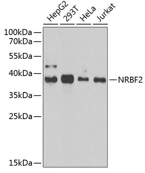
Western blot analysis of various lysates using NRBF2 Rabbit pAb (CAB6462) at 1:1000 dilution. Secondary antibody: HRP-conjugated Goat anti-Rabbit IgG (H+L) (CABS014) at 1:10000 dilution. Lysates/proteins: 25μg per lane. Blocking buffer: 3% nonfat dry milk in TBST. Detection: ECL Enhanced Kit (AbGn00021). Exposure time: 90s.
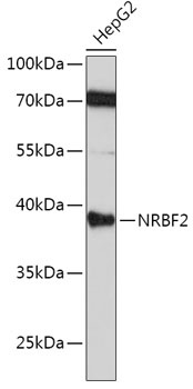
Western blot analysis of lysates from HepG2 cells, using [KO Validated] NRBF2 Rabbit pAb (CAB6462) at 1:1000 dilution. Secondary antibody: HRP-conjugated Goat anti-Rabbit IgG (H+L) (CABS014) at 1:10000 dilution. Lysates/proteins: 25μg per lane. Blocking buffer: 3% nonfat dry milk in TBST. Detection: ECL Basic Kit (AbGn00020). Exposure time: 3min.
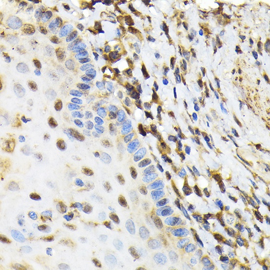
Immunohistochemistry analysis of paraffin-embedded Human esophageal using [KO Validated] NRBF2 Rabbit pAb (CAB6462) at dilution of 1:100 (40x lens). Microwave antigen retrieval performed with 0.01M Tris/EDTA Buffer (pH 9.0) prior to IHC staining.
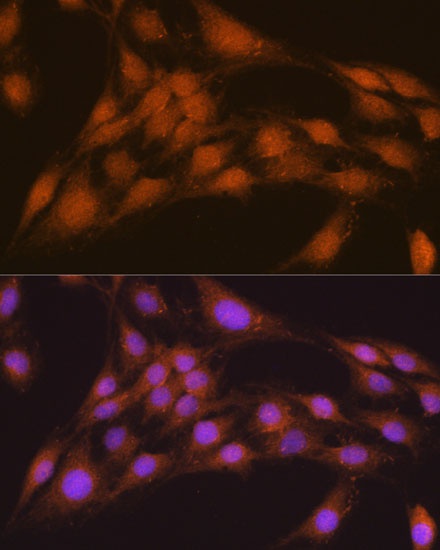
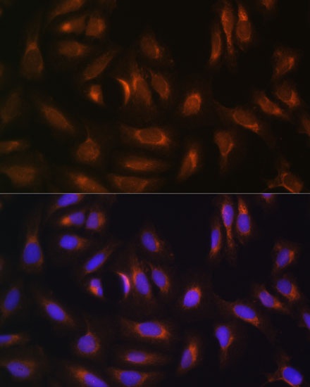
Immunofluorescence analysis of C6 cells using [KO Validated] NRBF2 Rabbit pAb (CAB6462) at dilution of 1:100. Secondary antibody: Cy3-conjugated Goat anti-Rabbit IgG (H+L) (CABS007) at 1:500 dilution. Blue: DAPI for nuclear staining.
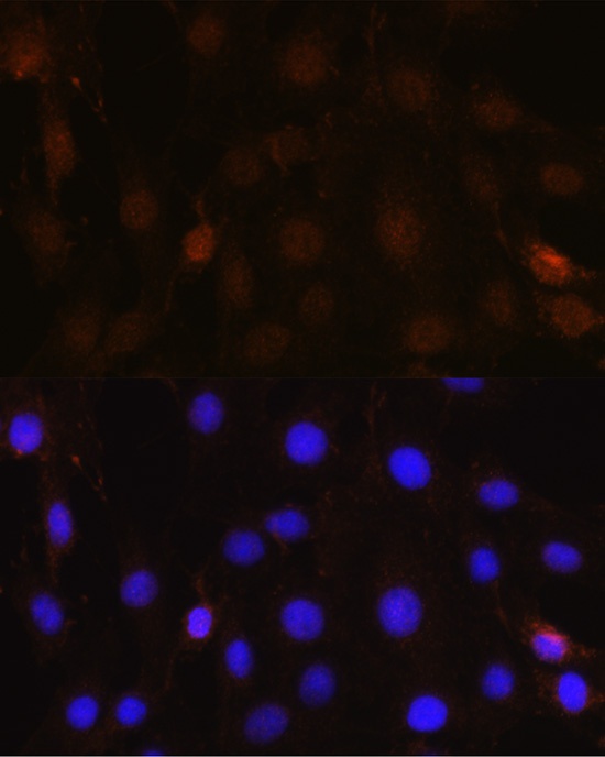
Immunofluorescence analysis of U-2 OS cells using [KO Validated] NRBF2 Rabbit pAb (CAB6462) at dilution of 1:100. Secondary antibody: Cy3-conjugated Goat anti-Rabbit IgG (H+L) (CABS007) at 1:500 dilution. Blue: DAPI for nuclear staining.
Immunofluorescence analysis of C6 cells using NRBF2 Rabbit pAb (CAB6462) at dilution of 1:100 (40x lens). Secondary antibody: Cy3-conjugated Goat anti-Rabbit IgG (H+L) (CABS007) at 1:500 dilution. Blue: DAPI for nuclear staining.
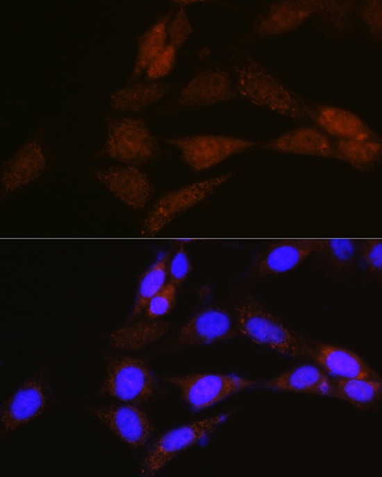
Immunofluorescence analysis of NIH-3T3 cells using NRBF2 Rabbit pAb (CAB6462) at dilution of 1:100 (40x lens). Secondary antibody: Cy3-conjugated Goat anti-Rabbit IgG (H+L) (CABS007) at 1:500 dilution. Blue: DAPI for nuclear staining.