The [KO Validated] NSDHL Antibody (CAB16893) is a high-quality antibody developed for reliable detection and analysis of target proteins. The antibody, produced in rabbits, exhibits high specificity for human samples and has been validated for use in Western blot applications. By targeting NSDHL, this antibody enables researchers to detect and analyze the protein in various cell types, making it ideal for studies in lipid metabolism, cholesterol homeostasis, and related diseases.NSDHL is a key enzyme in the mevalonate pathway, which plays a crucial role in the production of cholesterol, steroid hormones, and other important molecules in the body.
This antibody is validated for use in WB, IHC-P, IF/ICC, ELISA applications and has demonstrated reactivity against Human, Mouse, Rat samples.
Product Name:
[KO Validated] NSDHL Antibody
SKU:
CAB16893
Size:
20μL, 100μL
Reactivity:
Human, Mouse, Rat
Immunogen:
Recombinant protein (or fragment).This information is considered to be commercially sensitive.
Recommended starting concentration is 1 μg/mL. Please optimize the concentration based on your specific assay requirements.
Synonyms:
H105E3, XAP104, SDR31E1, HL
Positive Sample:
293T
Cellular Localization:
Endoplasmic Reticulum, Lipid Droplet.
Calculated MW:
42kDa
Observed MW:
40kDa
The protein encoded by this gene is localized in the endoplasmic reticulum and is involved in cholesterol biosynthesis. Mutations in this gene are associated with CHILD syndrome, which is a X-linked dominant disorder of lipid metabolism with disturbed cholesterol biosynthesis, and typically lethal in males. Alternatively spliced transcript variants with differing 5' UTR have been found for this gene.
Purification Method
Affinity purification
Gene ID
50814
RRID
AB_2770672
Buffer Information
Store at -20℃. Avoid freeze / thaw cycles. Buffer: PBS with 0.01% thimerosal,50% glycerol,pH7.3.
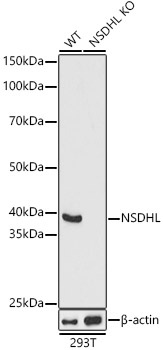
Western blot analysis of lysates from wild type(WT) and NSDHL knockout (KO) 293T(KO) cells, using [KO Validated] NSDHL Rabbit pAb (CAB16893) at 1:1000 dilution. Secondary antibody: HRP-conjugated Goat anti-Rabbit IgG (H+L) (CABS014) at 1:10000 dilution. Lysates/proteins: 25μg per lane. Blocking buffer: 3% nonfat dry milk in TBST. Detection: ECL Basic Kit (AbGn00020). Exposure time: 1s.
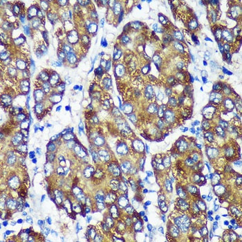
Immunohistochemistry analysis of paraffin-embedded Human liver cancer using [KO Validated] NSDHL Rabbit pAb (CAB16893) at dilution of 1:100 (40x lens). Microwave antigen retrieval performed with 0.01M PBS Buffer (pH 7.2) prior to IHC staining.
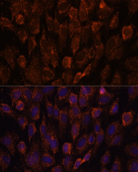
Immunofluorescence analysis of C6 cells using [KO Validated] NSDHL Rabbit pAb (CAB16893) at dilution of 1:100. Secondary antibody: Cy3-conjugated Goat anti-Rabbit IgG (H+L) (CABS007) at 1:500 dilution. Blue: DAPI for nuclear staining.
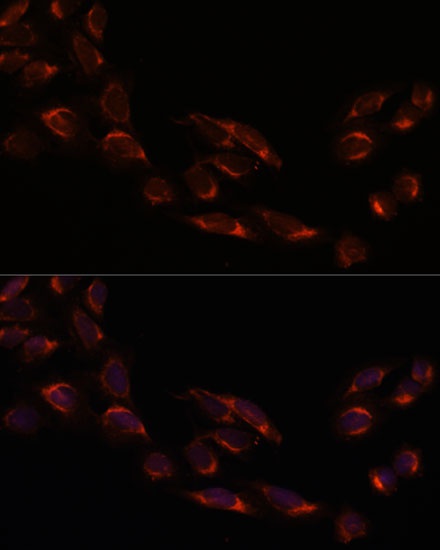
Immunofluorescence analysis of U-2 OS cells using [KO Validated] NSDHL Rabbit pAb (CAB16893) at dilution of 1:100. Secondary antibody: Cy3-conjugated Goat anti-Rabbit IgG (H+L) (CABS007) at 1:500 dilution. Blue: DAPI for nuclear staining.