The TrkB Antibody (CAB2099) is a high-quality antibody developed for reliable detection and analysis of target proteins. This antibody, produced in rabbits, has high reactivity with human samples and is suitable for use in Western blotting applications.NTRK2, also known as TrkB, is a key player in neurotrophic signaling pathways and has been implicated in various neurological and psychiatric disorders. By targeting the NTRK2 protein, this antibody allows for the detection and analysis of NTRK2 expression in different cell types, making it an ideal choice for studies in neuroscience and neurobiology.
This antibody is validated for use in WB, IF/ICC, ELISA applications and has demonstrated reactivity against Human, Mouse, Rat samples.
Product Name:
TrkB Antibody
SKU:
CAB2099
Size:
20μL, 100μL
Reactivity:
Human, Mouse, Rat
Conjugate:
Unconjugated
Immunogen:
Recombinant protein (or fragment).This information is considered to be commercially sensitive.
Cell Membrane, Endosome Membrane, Single-Pass Type I Membrane Protein.
Calculated MW:
92kDa
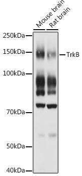
Observed MW:
140kDa
This gene encodes a member of the neurotrophic tyrosine receptor kinase (NTRK) family. This kinase is a membrane-bound receptor that, upon neurotrophin binding, phosphorylates itself and members of the MAPK pathway. Signalling through this kinase leads to cell differentiation. Mutations in this gene have been associated with obesity and mood disorders. Alternative splicing results in multiple transcript variants.
Purification Method
Affinity purification
Gene ID
4915
RRID
AB_2764118
Buffer Information
Store at -20℃. Avoid freeze / thaw cycles. Buffer: PBS containing 50% glycerol, preserved with proclin300 or sodium azide, pH 7.3.
Western blot analysis of various lysates using TrkB Rabbit pAb (CAB2099) at 1:1000 dilution. Secondary antibody: HRP-conjugated Goat anti-Rabbit IgG (H+L) (CABS014) at 1:10000 dilution. Lysates/proteins: 25μg per lane. Blocking buffer: 3% nonfat dry milk in TBST. Detection: ECL Basic Kit (AbGn00020). Exposure time: 30s.
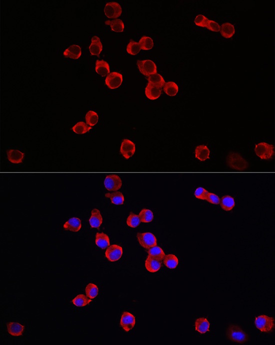
Immunofluorescence analysis of Neuro-2a cells using TrkB Rabbit pAb (CAB2099) at dilution of 1:100 (40x lens). Secondary antibody: Cy3-conjugated Goat anti-Rabbit IgG (H+L) (CABS007) at 1:500 dilution. Blue: DAPI for nuclear staining.