The NUDC Monoclonal Antibody (CAB4719) is a high-quality antibody developed for reliable detection and analysis of target proteins. This gene encodes a nuclear distribution protein that plays an essential role in mitosis and cytokinesis. The encoded protein is involved in spindle formation during mitosis and in microtubule organization during cytokinesis. Pseudogenes of this gene are found on chromosome 2.
This antibody is validated for use in WB, ELISA applications and has demonstrated reactivity against Human, Mouse, Rat samples.
Product Name:
NUDC Monoclonal Antibody
SKU:
CAB4719
Size:
100μL, 20μL
Reactivity:
Human, Mouse, Rat
Clone Number:
ARC1103
Conjugate:
Unconjugated
Immunogen:
Synthetic peptide. This information is considered to be commercially sensitive.
Tested Applications:
WBELISA
Recommended Dilution:
WB
1:500 - 1:2000
ELISA
Recommended starting concentration is 1 μg/mL. Please optimize the concentration based on your specific assay requirements.
Synonyms:
HNUDC, MNUDC, NPD011, NUDC
Positive Sample:
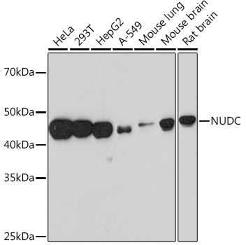
HeLa, 293T, HepG2, A-549, Mouse lung, Mouse brain, Rat brain
Cellular Localization:
Cytoplasm, Nucleus, Cytoskeleton.
Calculated MW:
38kDa
Observed MW:
42kDa
This gene encodes a nuclear distribution protein that plays an essential role in mitosis and cytokinesis. The encoded protein is involved in spindle formation during mitosis and in microtubule organization during cytokinesis. Pseudogenes of this gene are found on chromosome 2.
Purification Method
Affinity purification
Gene ID
10726
RRID
AB_2863330
Buffer Information
Store at -20℃. Avoid freeze / thaw cycles. Buffer: PBS containing 50% glycerol and 0.05% BSA, preserved with proclin300 or sodium azide, pH 7.3.
Western blot analysis of various lysates, using NUDC Rabbit mAb (CAB4719) at 1:1000 dilution. Secondary antibody: HRP-conjugated Goat anti-Rabbit IgG (H+L) (AS014) at 1:10000 dilution. Lysates/proteins: 25μg per lane. Blocking buffer: 3% nonfat dry milk in TBST. Detection: ECL Basic Kit (AbGn00020). Exposure time: 90s.