The OGDH Monoclonal Antibody (CAB22163) is a high-quality antibody developed for reliable detection and analysis of target proteins. This antibody, produced in mouse, has been rigorously validated for use in various applications, including immunohistochemistry and ELISA, and shows high specificity for OGDH protein detection.OGDH plays a vital role in cellular energy production and has been implicated in various diseases, including metabolic disorders and cancer.
This antibody is validated for use in WB, IHC-P, ELISA applications and has demonstrated reactivity against Human, Mouse, Rat samples.
Product Name:
OGDH Monoclonal Antibody
SKU:
CAB22163
Size:
20μL, 100μL
Reactivity:
Human, Mouse, Rat
Clone Number:
ARC55379
Conjugate:
Unconjugated
Immunogen:
Recombinant protein (or fragment).This information is considered to be commercially sensitive.
HeLa, MCF7, Mouse heart, Mouse skeletal muscle, Rat heart
Cellular Localization:
Mitochondrion, Nucleus.
Calculated MW:
116kDa
Observed MW:
116kDa
This gene encodes one subunit of the 2-oxoglutarate dehydrogenase complex. This complex catalyzes the overall conversion of 2-oxoglutarate (alpha-ketoglutarate) to succinyl-CoA and CO(2) during the Krebs cycle. The protein is located in the mitochondrial matrix and uses thiamine pyrophosphate as a cofactor. A congenital deficiency in 2-oxoglutarate dehydrogenase activity is believed to lead to hypotonia, metabolic acidosis, and hyperlactatemia. Alternative splicing results in multiple transcript variants encoding distinct isoforms.
Purification Method
Affinity purification
Gene ID
4967
Buffer Information
Store at -20℃. Avoid freeze / thaw cycles. Buffer: PBS with 0.09% Sodium azide,0.05% BSA,50% glycerol,pH7.3.
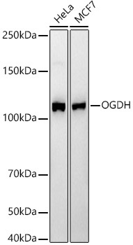
Western blot analysis of various lysates, using OGDH Rabbit mAb (CAB22163) at1:2000 dilution. Secondary antibody: HRP-conjugated Goat anti-Rabbit IgG (H+L) (CABS014) at 1:10000 dilution. Lysates/proteins: 25μg per lane. Blocking buffer: 3% nonfat dry milk in TBST. Detection: ECL Basic Kit (AbGn00020). Exposure time: 20s.
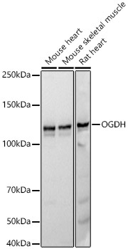
Western blot analysis of various lysates, using OGDH Rabbit mAb (CAB22163) at1:2000 dilution. Secondary antibody: HRP-conjugated Goat anti-Rabbit IgG (H+L) (CABS014) at 1:10000 dilution. Lysates/proteins: 25μg per lane. Blocking buffer: 3% nonfat dry milk in TBST. Detection: ECL Basic Kit (AbGn00020). Exposure time: 20s.
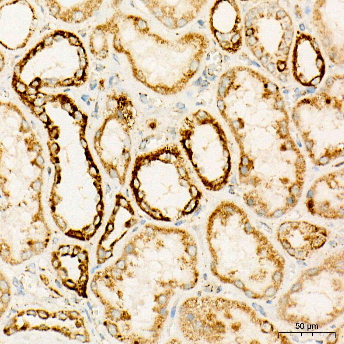
Immunohistochemistry analysis of paraffin-embedded Human kidney tissue using OGDH Rabbit mAb (CAB22163) at a dilution of 1:100 (40x lens). High pressure antigen retrieval performed with 0.01M Citrate buffer (pH 6.0) prior to IHC staining.
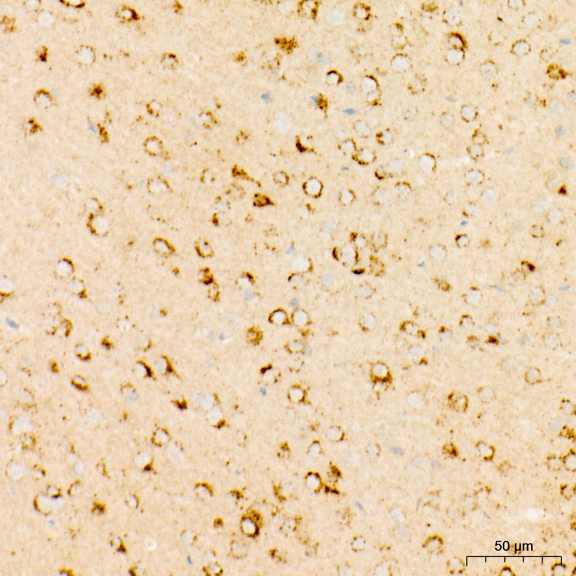
Immunohistochemistry analysis of paraffin-embedded Mouse brain tissue using OGDH Rabbit mAb (CAB22163) at a dilution of 1:100 (40x lens). High pressure antigen retrieval performed with 0.01M Citrate buffer (pH 6.0) prior to IHC staining.
 at1:2000 dilution. Secondary antibody: HRP Goat Anti-Rabbit IgG (H+L) at 1:10000 dilution. Lysates/proteins: 25μg per lane. Blocking buffer: 3% nonfat dry milk in TBST.")
 at1:2000 dilution. Secondary antibody: HRP Goat Anti-Rabbit IgG (H+L) at 1:10000 dilution. Lysates/proteins: 25μg per lane. Blocking buffer: 3% nonfat dry milk in TBST.")


