The p53 Polyclonal Antibody (CAB21630) is a high-quality antibody developed for reliable detection and analysis of target proteins. This antibody, generated in rabbits, exhibits high reactivity with human samples and has been extensively validated for use in Western blot applications.p53, also known as the "guardian of the genome," is involved in various cellular processes such as DNA repair, cell cycle control, and apoptosis. Mutations in the p53 gene are common in many types of cancer, underscoring the importance of studying its function and regulation.
This antibody is validated for use in WB, IHC-P, IF/ICC, IP, ELISA applications and has demonstrated reactivity against Human, Mouse samples.
Product Name:
p53 Polyclonal Antibody
SKU:
CAB21630
Size:
20μL, 100μL
Reactivity:
Human, Mouse
Conjugate:
Unconjugated
Immunogen:
Synthetic peptide. This information is considered to be commercially sensitive.
This gene encodes tumor protein p53, which responds to diverse cellular stresses to regulate target genes that induce cell cycle arrest, apoptosis, senescence, DNA repair, or changes in metabolism. p53 protein is expressed at low level in normal cells and at a high level in a variety of transformed cell lines, where it's believed to contribute to transformation and malignancy. p53 is a DNA-binding protein containing transcription activation, DNA-binding, and oligomerization domains. It is postulated to bind to a p53-binding site and activate expression of downstream genes that inhibit growth and/or invasion, and thus function as a tumor suppressor. Mice deficient for this gene are developmentally normal but are susceptible to spontaneous tumors. Evidence to date shows that this gene contains one promoter, in contrast to alternative promoters of the human gene, and transcribes a few of splice variants which encode different isoforms, although the biological validity or the full-length nature of some variants has not been determined.
Purification Method
Affinity purification
Gene ID
22059
Buffer Information
Store at -20℃. Avoid freeze / thaw cycles. Buffer: PBS containing 50% glycerol, preserved with proclin300 or sodium azide, pH 7.3.
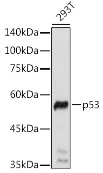
Western blot analysis of lysates from 293T cells, using p53 Rabbit pAb (CAB21630) at 1:1000 dilution. Secondary antibody: HRP-conjugated Goat anti-Rabbit IgG (H+L) (CABS014) at 1:10000 dilution. Lysates/proteins: 25μg per lane. Blocking buffer: 3% nonfat dry milk in TBST. Detection: ECL Basic Kit (AbGn00020). Exposure time: 5s.
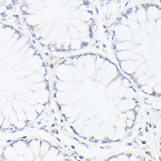
Immunohistochemistry analysis of paraffin-embedded Human colon using p53 Rabbit pAb (CAB21630) at dilution of 1:100 (40x lens). Microwave antigen retrieval performed with 0.01M PBS Buffer (pH 7.2) prior to IHC staining.
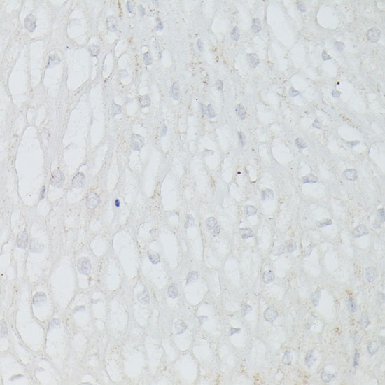
Immunohistochemistry analysis of paraffin-embedded Human esophageal using p53 Rabbit pAb (CAB21630) at dilution of 1:100 (40x lens). Microwave antigen retrieval performed with 0.01M PBS Buffer (pH 7.2) prior to IHC staining.
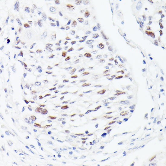
Immunohistochemistry analysis of paraffin-embedded Human esophageal cancer using p53 Rabbit pAb (CAB21630) at dilution of 1:100 (40x lens). Microwave antigen retrieval performed with 0.01M PBS Buffer (pH 7.2) prior to IHC staining.
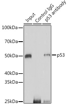
Immunoprecipitation analysis of 200 μg extracts of 293T cells using 3 μg p53 antibody (CAB21630). Western blot was performed from the immunoprecipitate using p53 antibody (CAB21630) at a dilution of 1:1000.
 at 1:1000 dilution. Secondary antibody: HRP Goat Anti-Rabbit IgG (H+L) at 1:10000 dilution. Lysates/proteins: 25μg per lane. Blocking buffer: 3% nonfat dry milk in TBST.")
 at 1:1000 dilution. Secondary antibody: HRP Goat Anti-Rabbit IgG (H+L) at 1:10000 dilution. Lysates/proteins: 25μg per lane. Blocking buffer: 3% nonfat dry milk in TBST.")


