The PABPC4 Antibody (CAB5948) is a high-quality antibody developed for reliable detection and analysis of target proteins. Poly(A)-binding proteins (PABPs) bind to the poly(A) tail present at the 3-prime ends of most eukaryotic mRNAs. PABPC4 or IPABP (inducible PABP) was isolated as an activation-induced T-cell mRNA encoding a protein. Activation of T cells increased PABPC4 mRNA levels in T cells approximately 5-fold. PABPC4 contains 4 RNA-binding domains and proline-rich C terminus. PABPC4 is localized primarily to the cytoplasm. It is suggested that PABPC4 might be necessary for regulation of stability of labile mRNA species in activated T cells. PABPC4 was also identified as an antigen, APP1 (activated-platelet protein-1), expressed on thrombin-activated rabbit platelets. PABPC4 may also be involved in the regulation of protein translation in platelets and megakaryocytes or may participate in the binding or stabilization of polyadenylates in platelet dense granules. Alternatively spliced transcript variants encoding different isoforms have been found for this gene. This protein has also been found to interact with coronavirus nucleocapsid proteins and is thought to inhibit coronavirus replication. RRID AB_2766680 Gene ID 8761 Swiss Prot Synonym APP1; APP-1; PABP4; iPABP; PABPC4
This antibody is validated for use in WB, IHC-P, IF/ICC, ELISA applications and has demonstrated reactivity against Human, Mouse, Rat samples.
Product Name:
PABPC4 Antibody
SKU:
CAB5948
Size:
100μL, 20μL
Reactivity:
Human, Mouse, Rat
Clone Number:
-
Conjugate:
Unconjugated
Immunogen:
Synthetic peptide. This information is considered to be commercially sensitive.
Tested Applications:
WBIHC-PIF/ICCELISA
Recommended Dilution:
WB
1:500 - 1:2000
IHC-P
1:50 - 1:200
IF
/
ICC
1:50 - 1:200
ELISA
Recommended starting concentration is 1 μg/mL. Please optimize the concentration based on your specific assay requirements.
Synonyms:
APP1, APP-1, PABP4, iPABP, PABPC4
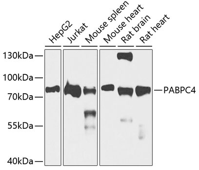
Positive Sample:
HepG2, Jurkat, Mouse spleen, Mouse heart, Rat brain, Rat heart
Cellular Localization:
Cytoplasm.
Calculated MW:
71kDa
Observed MW:
79kDa
Poly(A)-binding proteins (PABPs) bind to the poly(A) tail present at the 3-prime ends of most eukaryotic mRNAs. PABPC4 or IPABP (inducible PABP) was isolated as an activation-induced T-cell mRNA encoding a protein. Activation of T cells increased PABPC4 mRNA levels in T cells approximately 5-fold. PABPC4 contains 4 RNA-binding domains and proline-rich C terminus. PABPC4 is localized primarily to the cytoplasm. It is suggested that PABPC4 might be necessary for regulation of stability of labile mRNA species in activated T cells. PABPC4 was also identified as an antigen, APP1 (activated-platelet protein-1), expressed on thrombin-activated rabbit platelets. PABPC4 may also be involved in the regulation of protein translation in platelets and megakaryocytes or may participate in the binding or stabilization of polyadenylates in platelet dense granules. Alternatively spliced transcript variants encoding different isoforms have been found for this gene. This protein has also been found to interact with coronavirus nucleocapsid proteins and is thought to inhibit coronavirus replication. RRID AB_2766680 Gene ID 8761 Swiss Prot Synonym APP1; APP-1; PABP4; iPABP; PABPC4
Purification Method:
Affinity purification
Gene ID:
8761
RRID:
AB_2766680
Buffer Information:
Store at -20℃. Avoid freeze / thaw cycles. Buffer: PBS containing 50% glycerol, preserved with proclin300 or sodium azide, pH 7.3.
Western blot analysis of various lysates using PABPC4 Rabbit pAb (CAB5948) at 1:1000 dilution. Secondary antibody: HRP-conjugated Goat anti-Rabbit IgG (H+L) (AS014) at 1:10000 dilution. Lysates/proteins: 25μg per lane. Blocking buffer: 3% nonfat dry milk in TBST. Detection: ECL Basic Kit (AbGn00020). Exposure time: 30s.
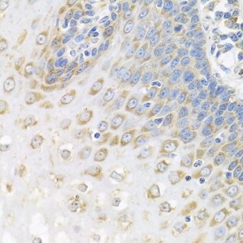
Immunohistochemistry analysis of paraffin-embedded Human esophagus using PABPC4 Rabbit pAb (CAB5948) at dilution of 1:100 (40x lens). Microwave antigen retrieval performed with 0.01M PBS Buffer (pH 7.2) prior to IHC staining.
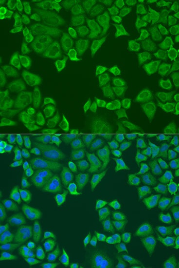
Immunofluorescence analysis of U2OS cells using PABPC4 Rabbit pAb (CAB5948) at dilution of 1:100. Secondary antibody: Cy3-conjugated Goat anti-Rabbit IgG (H+L) (AS007) at 1:500 dilution. Blue: DAPI for nuclear staining.