The PAH Antibody (CAB1559) is a high-quality antibody developed for reliable detection and analysis of target proteins. This antibody, produced in rabbits, exhibits high reactivity with human samples and is validated for use in Western blot applications. By binding to the PAH protein, this antibody enables accurate detection and analysis in various cell types, making it an ideal tool for studies in metabolic disorders and related research.PAH, also known as phenylalanine hydroxylase, plays a crucial role in the breakdown of phenylalanine, an essential amino acid. Mutations in the PAH gene can lead to phenylketonuria (PKU), a metabolic disorder that affects the body's ability to process phenylalanine.
This antibody is validated for use in WB, IF/ICC, ELISA applications and has demonstrated reactivity against Human, Mouse, Rat samples.
Product Name:
PAH Antibody
SKU:
CAB1559
Size:
20μL, 100μL
Reactivity:
Human, Mouse, Rat
Conjugate:
Unconjugated
Immunogen:
Recombinant protein (or fragment).This information is considered to be commercially sensitive.
Recommended starting concentration is 1 μg/mL. Please optimize the concentration based on your specific assay requirements.
Synonyms:
PH, PKU, PKU1, PAH
Positive Sample:
HepG2
Cellular Localization:
Cytosol.
Calculated MW:
52kDa
Observed MW:
52kDa
This gene encodes a member of the biopterin-dependent aromatic amino acid hydroxylase protein family. The encoded phenylalanine hydroxylase enzyme hydroxylates phenylalanine to tyrosine and is the rate-limiting step in phenylalanine catabolism. Deficiency of this enzyme activity results in the autosomal recessive disorder phenylketonuria.
Purification Method
Affinity purification
Gene ID
5053
RRID
AB_2762994
Buffer Information
Store at -20℃. Avoid freeze / thaw cycles. Buffer: PBS containing 50% glycerol, preserved with proclin300 or sodium azide, pH 7.3.
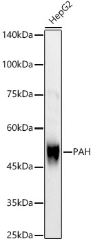
Western blot analysis of lysates from HepG2 cells, using PAH Rabbit pAb (CAB1559) at 1:700 dilution. Secondary antibody: HRP-conjugated Goat anti-Rabbit IgG (H+L) (CABS014) at 1:10000 dilution. Lysates/proteins: 25μg per lane. Blocking buffer: 3% nonfat dry milk in TBST. Detection: ECL Basic Kit (AbGn00020). Exposure time: 30s.
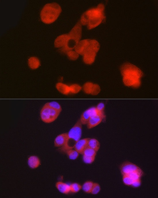
Immunofluorescence analysis of HepG2 cells using PAH Rabbit pAb (CAB1559) at dilution of 1:50 (40x lens). Secondary antibody: Cy3-conjugated Goat anti-Rabbit IgG (H+L) (CABS007) at 1:500 dilution. Blue: DAPI for nuclear staining.