The Pan Phospho-Tyrosine Antibody (CABP0905) is a high-quality antibody developed for reliable detection and analysis of target proteins. This antibody recognizes a wide range of proteins containing phosphorylated tyrosine residues, allowing for the detection and analysis of signaling pathways involved in various cellular processes.Phosphorylation events on tyrosine residues are crucial for the regulation of cell growth, differentiation, and survival. Abnormalities in tyrosine phosphorylation have been linked to diseases such as cancer, diabetes, and neurological disorders. The Pan-Phospho-Tyrosine Antibody enables researchers to investigate the role of tyrosine phosphorylation in these diseases and identify potential therapeutic targets.
This antibody is validated for use in WB, IF/ICC, ELISA applications and has demonstrated reactivity against Human, Mouse, Rat, Other (Wide Range Predicted) samples.
Product Name:
Pan Phospho-Tyrosine Antibody
SKU:
CABP0905
Size:
20μL, 100μL
Reactivity:
Human, Mouse, Rat, Other (Wide Range Predicted)
Immunogen:
Synthetic peptide. This information is considered to be commercially sensitive.
Tested Applications:
WBIF/ICCELISA
Recommended Dilution:
WB
1:500 - 1:2000
IF/ICC
1:50 - 1:200
ELISA
Recommended starting concentration is 1 μg/mL. Please optimize the concentration based on your specific assay requirements.
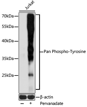
Positive Sample:
Jurkat treated with Pervanadate
Observed MW:
20-70kDa
Tyrosine phosphorylation (pTyr), much of which occurred on localized multiple sites, initiates cellular signaling, governs cellular functions, and its dysregulation is implicated in many diseases, especially cancers. pTyr-specific sensing is of great significance for understanding disease states and developing targeted anticancer drugs.
Purification Method
Affinity purification
RRID
AB_2770784
Buffer Information
Store at -20℃. Avoid freeze / thaw cycles. Buffer: PBS with 0.09% Sodium azide, 50% glycerol,pH7.3.
Western blot analysis of lysates from Jurkat cells, using Pan Phospho-Tyrosine pAb (CABP0905) at 1:1000 dilution. Jurkat cells were treated with Pervanadate (1 mM) at 37℃ for 30 minutes. Secondary antibody: HRP-conjugated Goat anti-Rabbit IgG (H+L) (CABS014) at 1:10000 dilution. Lysates/proteins: 25μg per lane. Blocking buffer: 3% BSA. Detection: ECL Basic Kit (AbGn00020). Exposure time: 1s.
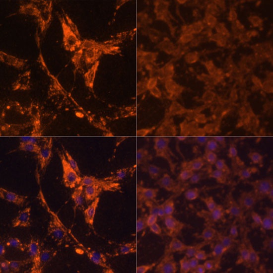
Immunofluorescence analysis of C6 cells using Pan Phospho-Tyrosine (CABP0905) at dilution of 1:100. Secondary antibody: Cy3-conjugated Goat anti-Rabbit IgG (H+L) (CABS007) at 1:500 dilution. Blue: DAPI for nuclear staining.C6 cells were treated with 20% FBS at 37℃ for 20 minutes after serum-starvation overnight.