The PARN Antibody (CAB6941) is a high-quality antibody developed for reliable detection and analysis of target proteins. This antibody, raised in rabbits, offers high sensitivity and specificity when detecting PARN in various experimental settings. Validated for use in Western blot applications, it provides researchers with a reliable method for analyzing PARN expression levels in different cell types.PARN is known for its role in mRNA degradation and processing, making it a crucial player in gene expression regulation. Dysregulation of PARN has been linked to various diseases, including cancer and neurological disorders, highlighting the importance of studying this enzyme in greater detail.
This antibody is validated for use in WB, IF/ICC, ELISA applications and has demonstrated reactivity against Human samples.
Product Name:
PARN Antibody
SKU:
CAB6941
Size:
20μL, 100μL
Reactivity:
Human
Conjugate:
Unconjugated
Immunogen:
Recombinant protein (or fragment).This information is considered to be commercially sensitive.
Recommended starting concentration is 1 μg/mL. Please optimize the concentration based on your specific assay requirements.
Synonyms:
DAN, DKCB6, PFBMFT4, PARN
Positive Sample:
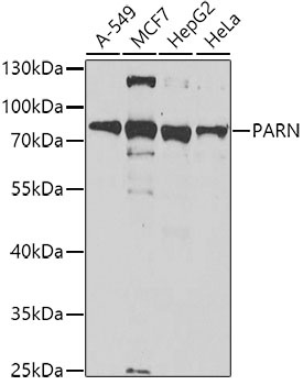
A-549, MCF7, HepG2, HeLa
Cellular Localization:
Cytoplasm, Nucleus, Nucleolus.
Calculated MW:
73kDa
Observed MW:
73kDa
The protein encoded by this gene is a 3'-exoribonuclease, with similarity to the RNase D family of 3'-exonucleases. It prefers poly(A) as the substrate, hence, efficiently degrades poly(A) tails of mRNAs. Exonucleolytic degradation of the poly(A) tail is often the first step in the decay of eukaryotic mRNAs. This protein is also involved in silencing of certain maternal mRNAs during oocyte maturation and early embryonic development, as well as in nonsense-mediated decay (NMD) of mRNAs that contain premature stop codons. Alternatively spliced transcript variants encoding different isoforms have been found for this gene.
Purification Method
Affinity purification
Gene ID
5073
RRID
AB_2767499
Buffer Information
Store at -20℃. Avoid freeze / thaw cycles. Buffer: PBS containing 50% glycerol, preserved with proclin300 or sodium azide, pH 7.3.
Western blot analysis of various lysates using PARN Rabbit pAb (CAB6941) at 1:1000 dilution. Secondary antibody: HRP-conjugated Goat anti-Rabbit IgG (H+L) (CABS014) at 1:10000 dilution. Lysates/proteins: 25μg per lane. Blocking buffer: 3% nonfat dry milk in TBST. Detection: ECL Basic Kit (AbGn00020). Exposure time: 150s.
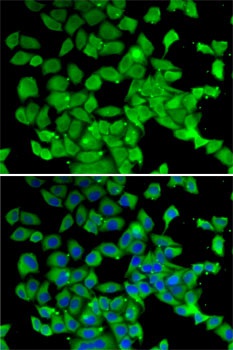
Immunofluorescence analysis of MCF7 cells using PARN Rabbit pAb (CAB6941). Secondary antibody: Cy3-conjugated Goat anti-Rabbit IgG (H+L) (CABS007) at 1:500 dilution. Blue: DAPI for nuclear staining.