The PAX2 Antibody (CAB3067) is a high-quality antibody developed for reliable detection and analysis of target proteins. PAX2 encodes paired box gene 2, one of many human homologues of the Drosophila melanogaster gene prd. The central feature of this transcription factor gene family is the conserved DNA-binding paired box domain. PAX2 is believed to be a target of transcriptional supression by the tumor suppressor gene WT1. Mutations within PAX2 have been shown to result in optic nerve colobomas and renal hypoplasia. Alternative splicing of this gene results in multiple transcript variants.
This antibody is validated for use in WB, IF/ICC, ELISA applications and has demonstrated reactivity against Human, Mouse, Rat samples.
Product Name:
PAX2 Antibody
SKU:
CAB3067
Size:
100μL, 20μL
Reactivity:
Human, Mouse, Rat
Conjugate:
Unconjugated
Immunogen:
Recombinant protein (or fragment).This information is considered to be commercially sensitive.
Tested Applications:
WBIF/ICCELISA
Recommended Dilution:
WB
1:100 - 1:500
IF/ICC
1:50 - 1:200
ELISA
Recommended starting concentration is 1 μg/mL. Please optimize the concentration based on your specific assay requirements.
Synonyms:
FSGS7, PAPRS, PAX-2, PAX2
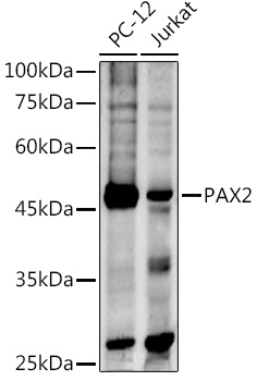
Positive Sample:
PC-12, Jurkat
Cellular Localization:
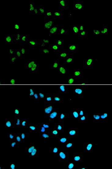
Nucleus.
Calculated MW:
45kDa
Observed MW:
47kDa
PAX2 encodes paired box gene 2, one of many human homologues of the Drosophila melanogaster gene prd. The central feature of this transcription factor gene family is the conserved DNA-binding paired box domain. PAX2 is believed to be a target of transcriptional supression by the tumor suppressor gene WT1. Mutations within PAX2 have been shown to result in optic nerve colobomas and renal hypoplasia. Alternative splicing of this gene results in multiple transcript variants.
Purification Method
Affinity purification
Gene ID
5076
RRID
AB_2764870
Buffer Information
Store at -20℃. Avoid freeze / thaw cycles. Buffer: PBS containing 50% glycerol, preserved with proclin300 or sodium azide, pH 7.3.
Western blot analysis of various lysates using PAX2 Rabbit pAb (CAB3067) at 1:500 dilution. Secondary antibody: HRP-conjugated Goat anti-Rabbit IgG (H+L) (AS014) at 1:10000 dilution. Lysates/proteins: 25μg per lane. Blocking buffer: 3% nonfat dry milk in TBST. Detection: ECL Enhanced Kit (AbGn00021). Exposure time: 180s.
Immunofluorescence analysis of A549 cells using PAX2 Rabbit pAb (CAB3067). Secondary antibody: Cy3-conjugated Goat anti-Rabbit IgG (H+L) (AS007) at 1:500 dilution. Blue: DAPI for nuclear staining.