The PBR/TSPO Monoclonal Antibody (CAB4881) is a high-quality antibody developed for reliable detection and analysis of target proteins. This antibody, produced in rabbits, is highly specific and suitable for use in Western blotting, immunofluorescence, and immunohistochemistry applications.The PBR protein, also known as Translocator Protein (TSPO), is involved in diverse cellular functions, including cholesterol transport, steroidogenesis, and immune response modulation. Its expression is often upregulated in conditions such as neuroinflammation, neurodegenerative diseases, and cancer, making it a potential biomarker and therapeutic target.
This antibody is validated for use in WB, IHC-P, IF/ICC, ELISA applications and has demonstrated reactivity against Human, Mouse samples.
Product Name:
PBR/TSPO Monoclonal Antibody
SKU:
CAB4881
Size:
20μL, 100μL
Reactivity:
Human, Mouse
Clone Number:
ARC0308
Conjugate:
Unconjugated
Immunogen:
Synthetic peptide. This information is considered to be commercially sensitive.
Present mainly in the mitochondrial compartment of peripheral tissues, the protein encoded by this gene interacts with some benzodiazepines and has different affinities than its endogenous counterpart. The protein is a key factor in the flow of cholesterol into mitochondria to permit the initiation of steroid hormone synthesis. Alternatively spliced transcript variants have been reported; one of the variants lacks an internal exon and is considered non-coding, and the other variants encode the same protein.
Purification Method
Affinity purification
Gene ID
706
RRID
AB_2863371
Buffer Information
Store at -20℃. Avoid freeze / thaw cycles. Buffer: PBS containing 50% glycerol and 0.05% BSA, preserved with proclin300 or sodium azide, pH 7.3.
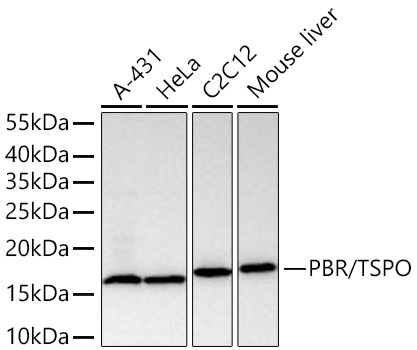
Western blot analysis of various lysates using PBR/TSPO Rabbit mAb (CAB4881) at 1:5000 dilution incubated overnight at 4℃. Secondary antibody: HRP-conjugated Goat anti-Rabbit IgG (H+L) (CABS014) at 1:10000 dilution. Lysates/proteins: 25 μg per lane. Blocking buffer: 3% nonfat dry milk in TBST. Detection: ECL Basic Kit (AbGn00020). Exposure time: 1s.
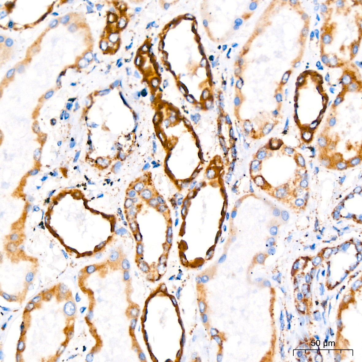
Immunohistochemistry analysis of paraffin-embedded Human kidney tissue using PBR/TSPO Rabbit mAb (CAB4881) at a dilution of 1:200 (40x lens). High pressure antigen retrieval performed with 0.01M Citrate Buffer (pH 6.0) prior to IHC staining.
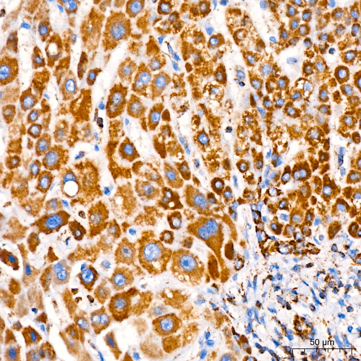
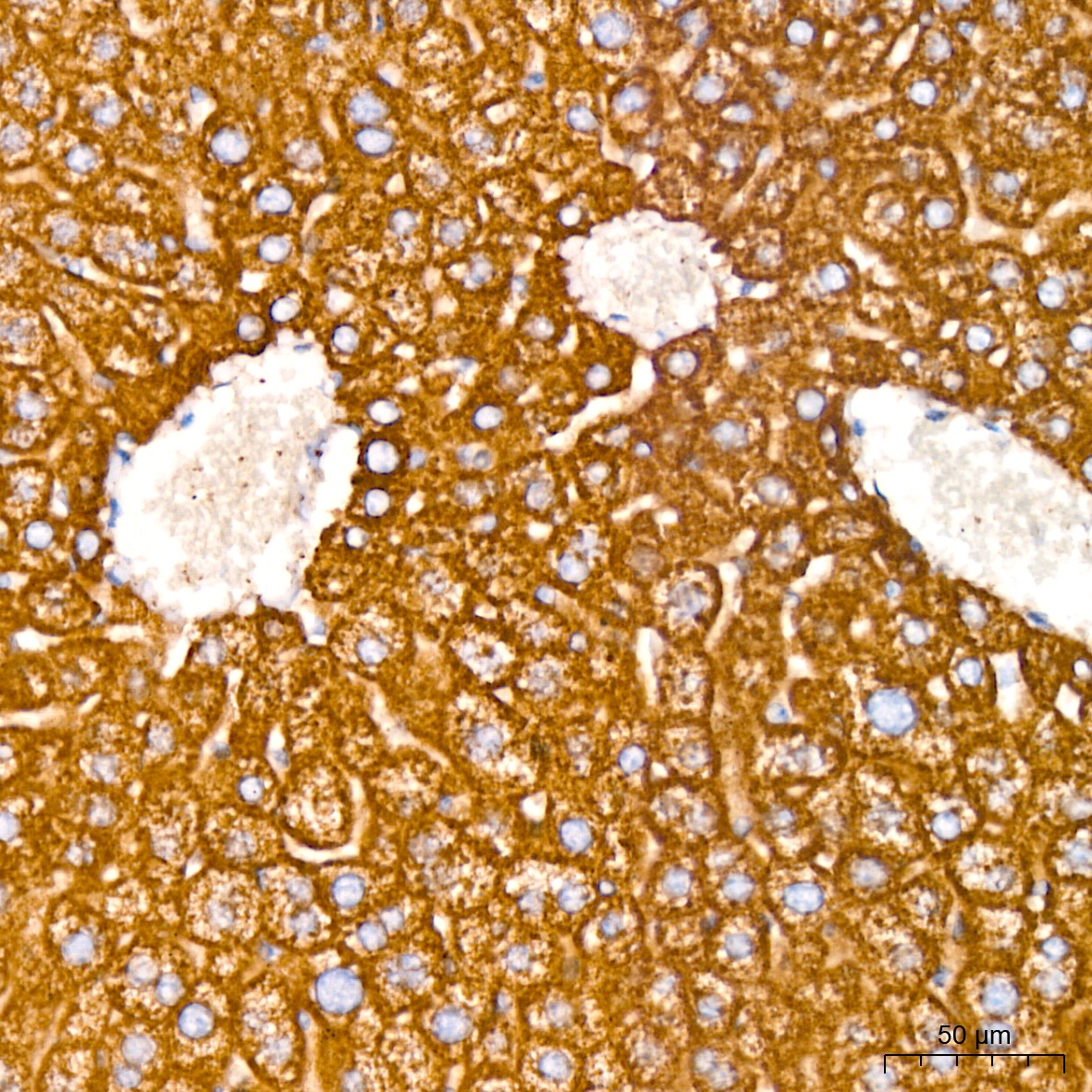
Immunohistochemistry analysis of paraffin-embedded Human liver tissue using PBR/TSPO Rabbit mAb (CAB4881) at a dilution of 1:200 (40x lens). High pressure antigen retrieval performed with 0.01M Citrate Buffer (pH 6.0) prior to IHC staining.
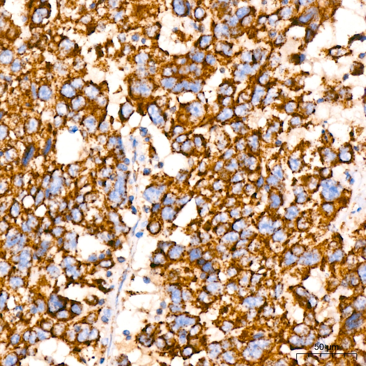
Immunohistochemistry analysis of paraffin-embedded Human lung cancer tissue using PBR/TSPO Rabbit mAb (CAB4881) at a dilution of 1:200 (40x lens). High pressure antigen retrieval performed with 0.01M Citrate Buffer (pH 6.0) prior to IHC staining.
Immunohistochemistry analysis of paraffin-embedded Mouse liver tissue using PBR/TSPO Rabbit mAb (CAB4881) at a dilution of 1:200 (40x lens). High pressure antigen retrieval performed with 0.01M Citrate Buffer (pH 6.0) prior to IHC staining.
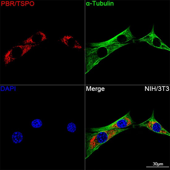
Confocal imaging of NIH/3T3 cells using PBR/TSPO Rabbit mAb (CAB4881,dilution 1:100)(Red). The cells were counterstained with α-Tubulin Mouse mAb (AC012,dilution 1:400) (Green). DAPI was used for nuclear staining (blue). Objective: 100x.
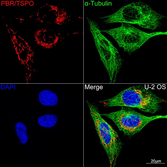
Confocal imaging of U-2 OS cells using PBR/TSPO Rabbit mAb (CAB4881,dilution 1:100)(Red). The cells were counterstained with α-Tubulin Mouse mAb (AC012,dilution 1:400) (Green). DAPI was used for nuclear staining (blue). Objective: 100x.