The PCBD1 Antibody (CAB6392) is a high-quality antibody developed for reliable detection and analysis of target proteins. This antibody, raised in rabbits, is specifically designed for use in Western blot applications and is highly reactive with human samples.PCBD1, also known as pterin-4-alpha-carbinolamine dehydratase 1, plays a crucial role in the tetrahydrobiopterin (BH4) pathway, which is essential for the production of neurotransmitters and nitric oxide. Dysregulation of this pathway has been implicated in various neurological disorders, making PCBD1 a target of interest in neuroscience research.
This antibody is validated for use in WB, IF/ICC, ELISA applications and has demonstrated reactivity against Human, Mouse, Rat samples.
Product Name:
PCBD1 Antibody
SKU:
CAB6392
Size:
20μL, 100μL
Reactivity:
Human, Mouse, Rat
Conjugate:
Unconjugated
Immunogen:
Recombinant protein (or fragment).This information is considered to be commercially sensitive.
Recommended starting concentration is 1 μg/mL. Please optimize the concentration based on your specific assay requirements.
Synonyms:
PCD, PHS, DCOH, PCBD, PCBD1
Positive Sample:
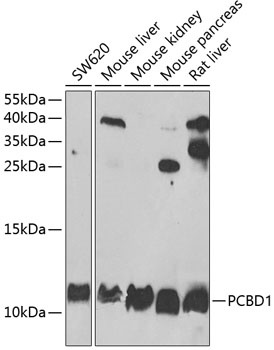
SW620, Mouse liver, Mouse kidney, Mouse pancreas, Rat liver
Cellular Localization:
Cytoplasm, Nucleus.
Calculated MW:
12kDa
Observed MW:
12kDa
This gene encodes a member of the pterin-4-alpha-carbinolamine dehydratase family. The encoded protein has been identified as a moonlighting protein based on its ability to perform mechanistically distinct functions. The encoded protein functions as both a dehydratase involved in tetrahydrobiopterin biosynthesis, and as a cofactor for HNF1A-dependent transcription. A deficiency of this enzyme leads to hyperphenylalaninemia. Alternative splicing results in multiple transcript variants.
Purification Method
Affinity purification
Gene ID
5092
RRID
AB_2766994
Buffer Information
Store at -20℃. Avoid freeze / thaw cycles. Buffer: PBS containing 50% glycerol, preserved with proclin300 or sodium azide, pH 7.3.
Western blot analysis of various lysates using PCBD1 Rabbit pAb (CAB6392) at 1:1000 dilution. Secondary antibody: HRP-conjugated Goat anti-Rabbit IgG (H+L) (CABS014) at 1:10000 dilution. Lysates/proteins: 25μg per lane. Blocking buffer: 3% nonfat dry milk in TBST. Detection: ECL Basic Kit (AbGn00020). Exposure time: 90s.
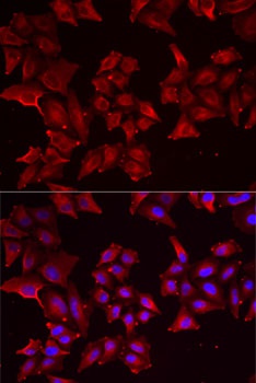
Immunofluorescence analysis of MCF7 cells using PCBD1 Rabbit pAb (CAB6392). Secondary antibody: Cy3-conjugated Goat anti-Rabbit IgG (H+L) (CABS007) at 1:500 dilution. Blue: DAPI for nuclear staining.