The PDAP1 Polyclonal Antibody (CAB4505) is a high-quality antibody developed for reliable detection and analysis of target proteins. This antibody, generated in rabbits, is highly specific to human samples and is validated for use in Western blot applications. It binds to the PDAP1 protein, allowing for accurate detection and analysis in a variety of cell types, making it an ideal choice for studies in molecular biology and biochemistry.PDAP1 is essential for the assembly of the mitochondrial ribosome and the translation of mitochondrial genes, making it a key player in cellular energy production and metabolism.
This antibody is validated for use in WB, IF/ICC, ELISA applications and has demonstrated reactivity against Human, Mouse, Rat samples.
Product Name:
PDAP1 Polyclonal Antibody
SKU:
CAB4505
Size:
20μL, 100μL
Reactivity:
Human, Mouse, Rat
Conjugate:
Unconjugated
Immunogen:
Synthetic peptide. This information is considered to be commercially sensitive.
Recommended starting concentration is 1 μg/mL. Please optimize the concentration based on your specific assay requirements.
Synonyms:
PAP, PAP1, HASPP28, PDAP1
Positive Sample:
MCF7, HeLa, NIH/3T3, Rat testis, Rat testis, MCF7, HeLa, NIH/3T3
Cellular Localization:
Cytosol, Extracellular Region, Plasma Membrane.
Calculated MW:
21kDa
Observed MW:
26kDa/25kDa
The protein encoded by this gene is a phosphoprotein that may upregulate the PDGFA-stimulated growth of fibroblasts and also downregulate the mitogenicity of PDGFB. The encoded protein in rodents has been shown to bind PDGFA with a low affinity.
Purification Method
Affinity purification
Gene ID
11333
RRID
AB_2765721
Buffer Information
Store at -20℃. Avoid freeze / thaw cycles. Buffer: PBS containing 50% glycerol, preserved with proclin300 or sodium azide, pH 7.3.
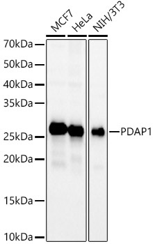
Western blot analysis of various lysates using PDAP1 Rabbit pAb (CAB4505) at 1:500 dilution. Secondary antibody: HRP-conjugated Goat anti-Rabbit IgG (H+L) (CABS014) at 1:10000 dilution. Lysates / proteins: 25 μg per lane. Blocking buffer: 3 % nonfat dry milk in TBST. Detection: ECL Basic Kit (AbGn00020). Exposure time: 30s.
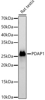
Western blot analysis of lysates from Rat testis using PDAP1 Rabbit pAb (CAB4505) at 1:500 dilution. Secondary antibody: HRP-conjugated Goat anti-Rabbit IgG (H+L) (CABS014) at 1:10000 dilution. Lysates/proteins: 25 μg per lane. Blocking buffer: 3% nonfat dry milk in TBST. Detection: ECL Basic Kit (AbGn00020). Exposure time: 60s.
 at dilution of 1:50 (40x lens). Secondary antibody: Cy3 Goat Anti-Rabbit IgG (H+L) (AS007) at 1:500 dilution. Blue: DAPI for nuclear staining.")
 at dilution of 1:50 (40x lens). Secondary antibody: Cy3 Goat Anti-Rabbit IgG (H+L) (AS007) at 1:500 dilution. Blue: DAPI for nuclear staining.")

")
