The PDC Antibody (CAB14654) is a high-quality antibody developed for reliable detection and analysis of target proteins. This antibody, produced in rabbits, exhibits high reactivity with human samples and is specifically validated for use in Western blot applications.PDC, also known as plasmacytoid dendritic cell antigen, plays a pivotal role in antiviral immunity and the activation of immune responses. Its expression has been linked to various inflammatory diseases, making it a promising target for therapeutic development and disease research.
This antibody is validated for use in WB, IHC-P, ELISA applications and has demonstrated reactivity against Mouse, Rat samples.
Product Name:
PDC Antibody
SKU:
CAB14654
Size:
20μL, 100μL
Reactivity:
Mouse, Rat
Conjugate:
Unconjugated
Immunogen:
Recombinant protein (or fragment).This information is considered to be commercially sensitive.
This gene encodes a phosphoprotein, which is located in the outer and inner segments of the rod cells in the retina. This protein may participate in the regulation of visual phototransduction or in the integration of photoreceptor metabolism. It modulates the phototransduction cascade by interacting with the beta and gamma subunits of the retinal G-protein transducin. This gene is a potential candidate gene for retinitis pigmentosa and Usher syndrome type II. Alternatively spliced transcript variants encoding different isoforms have been identified.
Purification Method
Affinity purification
Gene ID
5132
RRID
AB_2761530
Buffer Information
Store at -20℃. Avoid freeze / thaw cycles. Buffer: PBS containing 50% glycerol, preserved with proclin300 or sodium azide, pH 7.3.
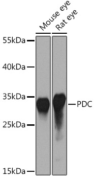
Western blot analysis of various lysates using PDC Rabbit pAb (CAB14654) at 1:1000 dilution. Secondary antibody: HRP-conjugated Goat anti-Rabbit IgG (H+L) (CABS014) at 1:10000 dilution. Lysates/proteins: 25μg per lane. Blocking buffer: 3% nonfat dry milk in TBST. Detection: ECL Basic Kit (AbGn00020). Exposure time: 10s.
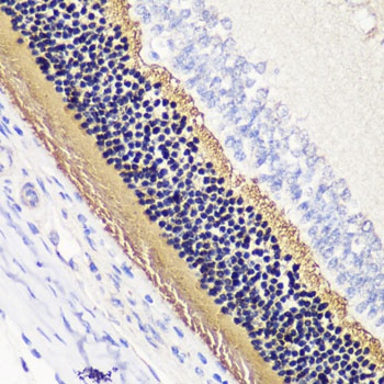
Immunohistochemistry analysis of paraffin-embedded Rat retina using PDC Rabbit pAb (CAB14654) at dilution of 1:200 (40x lens). Microwave antigen retrieval performed with 0.01M PBS Buffer (pH 7.2) prior to IHC staining.