The PDE4B Polyclonal Antibody (CAB23257) is a high-quality antibody developed for reliable detection and analysis of target proteins. The antibody, produced in rabbits, exhibits high reactivity with human samples and is validated for use in Western blot applications. It specifically binds to the PDE4B protein, allowing for accurate detection and analysis in a wide range of cell types.PDE4B is a key player in intracellular signaling pathways that control inflammation and immune cell function. Dysregulation of PDE4B has been linked to several inflammatory diseases, making it a potential therapeutic target for conditions such as asthma, COPD, and rheumatoid arthritis.
This antibody is validated for use in WB, ELISA applications and has demonstrated reactivity against Mouse, Rat samples.
Product Name:
PDE4B Polyclonal Antibody
SKU:
CAB23257
Size:
20μL, 100μL
Reactivity:
Mouse, Rat
Conjugate:
Unconjugated
Immunogen:
Recombinant protein (or fragment).This information is considered to be commercially sensitive.
Recommended starting concentration is 1 μg/mL. Please optimize the concentration based on your specific assay requirements.
Synonyms:
DPDE4, PDEIVB, PDE4B
Positive Sample:
Mouse lung
Cellular Localization:
Centrosome, Cytosol, Dendritic Spine, Excitatory Synapse, Gamma-Tubulin Complex, Nucleus, Perinuclear Region Of Cytoplasm, Postsynaptic Density, Synaptic Vesicle, Z Disc.
Calculated MW:
83kDa
Observed MW:
90kDa
This gene is a member of the type IV, cyclic AMP (cAMP)-specific, cyclic nucleotide phosphodiesterase (PDE) family. The encoded protein regulates the cellular concentrations of cyclic nucleotides and thereby play a role in signal transduction. Altered activity of this protein has been associated with schizophrenia and bipolar affective disorder. Alternative splicing and the use of alternative promoters results in multiple transcript variants encoding different isoforms.
Purification Method
Affinity purification
Gene ID
5142
Buffer Information
Store at -20℃. Avoid freeze / thaw cycles. Buffer: PBS containing 50% glycerol, preserved with proclin300 or sodium azide, pH 7.3.
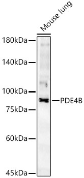
Western blot analysis of lysates from Mouse lung, using PDE4B Rabbit pAb (CAB23257) at 1:2000 dilution. Secondary antibody: HRP-conjugated Goat anti-Rabbit IgG (H+L) (CABS014) at 1:10000 dilution. Lysates/proteins: 25μg per lane. Blocking buffer: 3% nonfat dry milk in TBST. Detection: ECL Basic Kit (AbGn00020). Exposure time: 180s.
 at 1:2000 dilution. Secondary antibody: HRP Goat Anti-Rabbit IgG (H+L) at 1:10000 dilution. Lysates/proteins: 25μg per lane. Blocking buffer: 3% nonfat dry milk in TBST.")
 at 1:2000 dilution. Secondary antibody: HRP Goat Anti-Rabbit IgG (H+L) at 1:10000 dilution. Lysates/proteins: 25μg per lane. Blocking buffer: 3% nonfat dry milk in TBST.")


")
 at 1:10000 dilution. Lysates/proteins: 25ug per lane. Blocking buffer: 3% nonfat dry milk in TBST. Detection: ECL Basic Kit. Exposure time: 1s.")