The PDGFD Antibody (CAB14653) is a high-quality antibody developed for reliable detection and analysis of target proteins. This antibody, produced in rabbits, exhibits high specificity and robust reactivity with human samples, making it an ideal choice for Western blot applications.PDGFD is a key player in the regulation of cell proliferation, migration, and survival, making it an attractive target for investigations in developmental biology, angiogenesis, and tissue repair. The PDGFD Polyclonal Antibody binds specifically to the PDGFD protein, allowing for precise detection and analysis in a variety of cell types and experimental settings.
This antibody is validated for use in WB, ELISA applications and has demonstrated reactivity against Human, Mouse, Rat samples.
Product Name:
PDGFD Antibody
SKU:
CAB14653
Size:
20μL, 100μL
Reactivity:
Human, Mouse, Rat
Conjugate:
Unconjugated
Immunogen:
Synthetic peptide. This information is considered to be commercially sensitive.
Recommended starting concentration is 1 μg/mL. Please optimize the concentration based on your specific assay requirements.
Synonyms:
IEGF, SCDGFB, MSTP036, SCDGF-B, PDGFD
Positive Sample:
HeLa, Mouse heart, Rat kidney
Cellular Localization:
Secreted.
Calculated MW:
43kDa
Observed MW:
50kDa
The protein encoded by this gene is a member of the platelet-derived growth factor family. The four members of this family are mitogenic factors for cells of mesenchymal origin and are characterized by a core motif of eight cysteines, seven of which are found in this factor. This gene product only forms homodimers and, therefore, does not dimerize with the other three family members. It differs from alpha and beta members of this family in having an unusual N-terminal domain, the CUB domain. Two splice variants have been identified for this gene.
Purification Method
Affinity purification
Gene ID
80310
RRID
AB_2761529
Buffer Information
Store at -20℃. Avoid freeze / thaw cycles. Buffer: PBS containing 50% glycerol, preserved with proclin300 or sodium azide, pH 7.3.
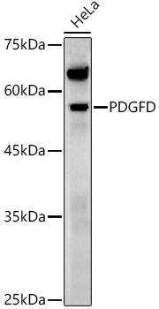
Western blot analysis of various lysates using PDGFD Rabbit pAb (CAB14653) at 1:1000 dilution. Secondary antibody: HRP-conjugated Goat anti-Rabbit IgG (H+L) (CABS014) at 1:10000 dilution. Lysates/proteins: 25μg per lane. Blocking buffer: 3% nonfat dry milk in TBST. Detection: ECL Enhanced Kit (AbGn00021). Exposure time: 60s.
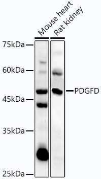
Western blot analysis of various lysates using PDGFD Rabbit pAb (CAB14653) at 1:1000 dilution. Secondary antibody: HRP-conjugated Goat anti-Rabbit IgG (H+L) (CABS014) at 1:10000 dilution. Lysates/proteins: 25μg per lane. Blocking buffer: 3% nonfat dry milk in TBST. Detection: ECL Enhanced Kit (AbGn00021). Exposure time: 30s.