The PDHB Monoclonal Antibody (CAB4645) is a high-quality antibody developed for reliable detection and analysis of target proteins. This monoclonal antibody, developed using rabbit-derived cells, is highly specific and reactive to human PDHB samples, making it ideal for a variety of research applications.PDHB is a critical component of the pyruvate dehydrogenase complex, which plays a key role in cellular metabolism and energy production. Dysregulation of PDHB has been linked to various diseases, including cancer, diabetes, and neurodegenerative disorders. The PDHB Rabbit Monoclonal Antibody enables precise detection and analysis of PDHB in different cell types, providing researchers with a reliable tool for exploring its function and potential therapeutic targets.
This antibody is validated for use in WB, IHC-P, ELISA applications and has demonstrated reactivity against Human, Mouse, Rat samples.
Product Name:
PDHB Monoclonal Antibody
SKU:
CAB4645
Size:
20μL, 100μL
Reactivity:
Human, Mouse, Rat
Clone Number:
ARC1074
Conjugate:
Unconjugated
Immunogen:
Synthetic peptide. This information is considered to be commercially sensitive.
Recommended starting concentration is 1 μg/mL. Please optimize the concentration based on your specific assay requirements.
Synonyms:
PDHBD, PHE1B, E1beta, PDHE1B, PDHE1-B, PDHB
Positive Sample:
293T, K-562, U-251MG, Mouse liver, Mouse skeletal muscle, Mouse heart, Rat skeletal muscle, Rat kidney, Rat heart
Cellular Localization:
Mitochondrion Matrix.
Calculated MW:
39kDa
Observed MW:
35-39kDa
The pyruvate dehydrogenase (PDH) complex is a nuclear-encoded mitochondrial multienzyme complex that catalyzes the overall conversion of pyruvate to acetyl-CoA and carbon dioxide, and provides the primary link between glycolysis and the tricarboxylic acid (TCA) cycle. The PDH complex is composed of multiple copies of three enzymatic components: pyruvate dehydrogenase (E1), dihydrolipoamide acetyltransferase (E2) and lipoamide dehydrogenase (E3). The E1 enzyme is a heterotetramer of two alpha and two beta subunits. This gene encodes the E1 beta subunit. Mutations in this gene are associated with pyruvate dehydrogenase E1-beta deficiency. Alternatively spliced transcript variants have been found for this gene.
Purification Method
Affinity purification
Gene ID
5162
RRID
AB_2863316
Buffer Information
Store at -20℃. Avoid freeze / thaw cycles. Buffer: PBS containing 50% glycerol and 0.05% BSA, preserved with proclin300 or sodium azide, pH 7.3.
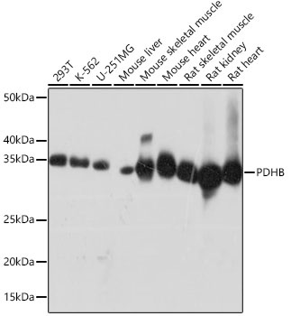
Western blot analysis of various lysates using PDHB Rabbit mAb (CAB4645)at 1:1000 dilution. Secondary antibody: HRP-conjugated Goat anti-Rabbit IgG (H+L) (CABS014) at 1:10000 dilution. Lysates/proteins: 25 μg per lane. Blocking buffer: 3% nonfat dry milk in TBST. Detection: ECL Basic Kit (AbGn00020). Exposure time: 10s.
Immunohistochemistry analysis of paraffin-embedded Human thyroid cancer tissue using PDHB Rabbit mAb (CAB4645) at a dilution of 1:200 (40x lens). High pressure antigen retrieval was performed with 0.01 M citrate buffer (pH 6.0) prior to IHC staining.
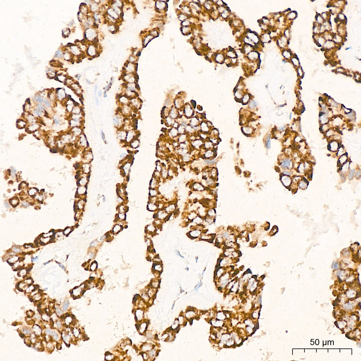
Immunohistochemistry analysis of paraffin-embedded Human lung cancer tissue using PDHB Rabbit mAb (CAB4645) at a dilution of 1:200 (40x lens). High pressure antigen retrieval was performed with 0.01 M citrate buffer (pH 6.0) prior to IHC staining.
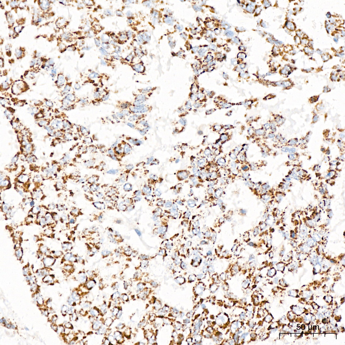
Immunohistochemistry analysis of paraffin-embedded Human colon carcinoma tissue using PDHB Rabbit mAb (CAB4645) at a dilution of 1:200 (40x lens). High pressure antigen retrieval was performed with 0.01 M citrate buffer (pH 6.0) prior to IHC staining.
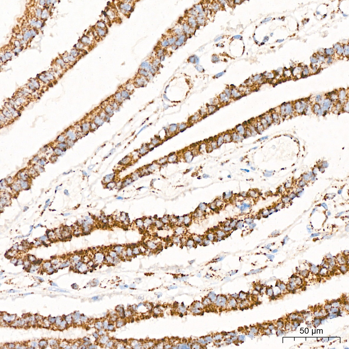
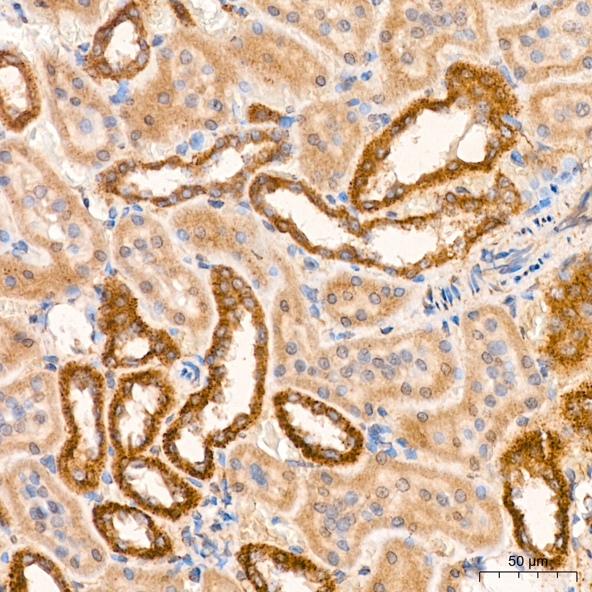
Immunohistochemistry analysis of paraffin-embedded Rat kidney tissue using PDHB Rabbit mAb (CAB4645) at a dilution of 1:200 (40x lens). High pressure antigen retrieval was performed with 0.01 M citrate buffer (pH 6.0) prior to IHC staining.