The PDHX Antibody (CAB6426) is a high-quality antibody developed for reliable detection and analysis of target proteins. This antibody, produced in rabbits, is highly sensitive and specific for detecting PDHX protein in human samples, making it ideal for Western blot analysis.PDHX is a crucial component of the PDC, playing a critical role in the conversion of pyruvate to acetyl-CoA, a fundamental step in cellular energy metabolism.
This antibody is validated for use in WB, IP, ELISA applications and has demonstrated reactivity against Human, Mouse, Rat samples.
Product Name:
PDHX Antibody
SKU:
CAB6426
Size:
20μL, 100μL
Reactivity:
Human, Mouse, Rat
Conjugate:
Unconjugated
Immunogen:
Recombinant protein (or fragment).This information is considered to be commercially sensitive.
0.5μg-4μg antibody for 400μg-600μg extracts of whole cells
ELISA
Recommended starting concentration is 1 μg/mL. Please optimize the concentration based on your specific assay requirements.
Synonyms:
E3BP, OPDX, PDX1, proX, DLDBP, PDHXD, PDHX
Positive Sample:
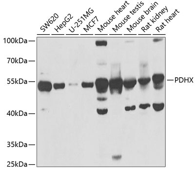
SW620, HepG2, U-251MG, MCF7, Mouse heart, Mouse testis, Mouse brain, Rat kidney, Rat heart
Cellular Localization:
Mitochondrion Matrix.
Calculated MW:
54kDa
Observed MW:
54kDa
The pyruvate dehydrogenase (PDH) complex is located in the mitochondrial matrix and catalyzes the conversion of pyruvate to acetyl coenzyme A. The PDH complex thereby links glycolysis to Krebs cycle. The PDH complex contains three catalytic subunits, E1, E2, and E3, two regulatory subunits, E1 kinase and E1 phosphatase, and a non-catalytic subunit, E3 binding protein (E3BP). This gene encodes the E3 binding protein subunit; also known as component X of the pyruvate dehydrogenase complex. This protein tethers E3 dimers to the E2 core of the PDH complex. Defects in this gene are a cause of pyruvate dehydrogenase deficiency which results in neurological dysfunction and lactic acidosis in infancy and early childhood. This protein is also a minor antigen for antimitochondrial antibodies. These autoantibodies are present in nearly 95% of patients with the autoimmune liver disease primary biliary cirrhosis (PBC). In PBC, activated T lymphocytes attack and destroy epithelial cells in the bile duct where this protein is abnormally distributed and overexpressed. PBC eventually leads to cirrhosis and liver failure. Alternative splicing results in multiple transcript variants encoding distinct isoforms.
Purification Method
Affinity purification
Gene ID
8050
RRID
AB_2767028
Buffer Information
Store at -20℃. Avoid freeze / thaw cycles. Buffer: PBS containing 50% glycerol, preserved with proclin300 or sodium azide, pH 7.3.
Western blot analysis of various lysates using PDHX Rabbit pAb (CAB6426) at 1:1000 dilution. Secondary antibody: HRP-conjugated Goat anti-Rabbit IgG (H+L) (CABS014) at 1:10000 dilution. Lysates/proteins: 25μg per lane. Blocking buffer: 3% nonfat dry milk in TBST. Detection: ECL Basic Kit (AbGn00020). Exposure time: 5s.
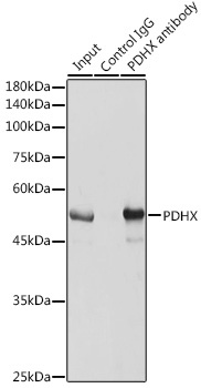
Immunoprecipitation analysis of 600 μg extracts of Mouse brain cells using 3 μg PDHX antibody (CAB6426). Western blot was performed from the immunoprecipitate using PDHX antibody (CAB6426) at a dilution of 1:1000.