The PDP1 Antibody (CAB14545) is a high-quality antibody developed for reliable detection and analysis of target proteins. This antibody, produced in rabbits, is highly specific for human samples and is validated for use in applications such as Western blotting. By binding to the PDP1 protein, this antibody allows for the detection and analysis of PDP1 in various cell types, making it ideal for studies in the fields of biochemistry and cellular metabolism.PDP1, also known as pyruvate dehyrogenase phosphatase subunit 1, is a crucial enzyme involved in the regulation of glucose metabolism and energy production within cells.
This antibody is validated for use in WB, IF/ICC, ELISA applications and has demonstrated reactivity against Human, Mouse, Rat samples.
Product Name:
PDP1 Antibody
SKU:
CAB14545
Size:
20μL, 100μL
Reactivity:
Human, Mouse, Rat
Conjugate:
Unconjugated
Immunogen:
Recombinant protein (or fragment).This information is considered to be commercially sensitive.
Recommended starting concentration is 1 μg/mL. Please optimize the concentration based on your specific assay requirements.
Synonyms:
PDH, PDP, PDPC, PPM2A, PPM2C, PDP1
Positive Sample:
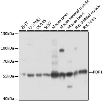
293T, U-87MG, DU145, 5637, mouse brain, mouse skeletal muscle, mouse heart, rat skeletal muscle, rat heart
Cellular Localization:
Mitochondrion Matrix.
Calculated MW:
61kDa
Observed MW:
61kDa
Pyruvate dehydrogenase (E1) is one of the three components (E1, E2, and E3) of the large pyruvate dehydrogenase complex. Pyruvate dehydrogenase kinases catalyze phosphorylation of serine residues of E1 to inactivate the E1 component and inhibit the complex. Pyruvate dehydrogenase phosphatases catalyze the dephosphorylation and activation of the E1 component to reverse the effects of pyruvate dehydrogenase kinases. Pyruvate dehydrogenase phosphatase is a heterodimer consisting of catalytic and regulatory subunits. Two catalytic subunits have been reported; one is predominantly expressed in skeletal muscle and another one is is much more abundant in the liver. The catalytic subunit, encoded by this gene, is the former, and belongs to the protein phosphatase 2C (PP2C) superfamily. Along with the pyruvate dehydrogenase complex and pyruvate dehydrogenase kinases, this enzyme is located in the mitochondrial matrix. Mutation in this gene causes pyruvate dehydrogenase phosphatase deficiency. Multiple alternatively spliced transcript variants encoding different isoforms have been identified.
Purification Method
Affinity purification
Gene ID
54704
RRID
AB_2761421
Buffer Information
Store at -20℃. Avoid freeze / thaw cycles. Buffer: PBS with 0.01% thimerosal,50% glycerol,pH7.3.
Western blot analysis of various lysates using PDP1 Rabbit pAb (CAB14545) at 1:3000 dilution. Secondary antibody: HRP-conjugated Goat anti-Rabbit IgG (H+L) (CABS014) at 1:10000 dilution. Lysates/proteins: 25μg per lane. Blocking buffer: 3% nonfat dry milk in TBST. Detection: ECL Basic Kit (AbGn00020). Exposure time: 1s.
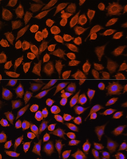
Immunofluorescence analysis of L929 cells using PDP1 Rabbit pAb (CAB14545) at dilution of 1:100. Secondary antibody: Cy3-conjugated Goat anti-Rabbit IgG (H+L) (CABS007) at 1:500 dilution. Blue: DAPI for nuclear staining.