The PEPCK/PCK2 Polyclonal Antibody (CAB21529) is a high-quality antibody developed for reliable detection and analysis of target proteins. This antibody is raised in rabbits and has been validated for use in Western blot applications, allowing for the detection and analysis of PEPCK and PCK2 in a variety of cell types.PEPCK and PCK2 are key enzymes involved in gluconeogenesis, the process by which glucose is synthesized from non-carbohydrate sources. These enzymes play a crucial role in regulating blood sugar levels and are therefore of interest in studies of diabetes, obesity, and metabolic disorders.
This antibody is validated for use in WB, IHC-P, IF/ICC, IP, ELISA applications and has demonstrated reactivity against Human, Mouse, Rat samples.
Product Name:
PEPCK/PCK2 Polyclonal Antibody
SKU:
CAB21529
Size:
20μL, 100μL
Reactivity:
Human, Mouse, Rat
Conjugate:
Unconjugated
Immunogen:
Recombinant protein (or fragment).This information is considered to be commercially sensitive.
0.5μg-4μg antibody for 200μg-400μg extracts of whole cells
ELISA
Recommended starting concentration is 1 μg/mL. Please optimize the concentration based on your specific assay requirements.
Synonyms:
PEPCK, PEPCK2, PEPCK-M, K2
Positive Sample:
293T, SH-SY5Y, HepG2
Cellular Localization:
Mitochondrion.
Calculated MW:
71kDa
Observed MW:
71kDa
This gene encodes a mitochondrial enzyme that catalyzes the conversion of oxaloacetate to phosphoenolpyruvate in the presence of guanosine triphosphate (GTP). A cytosolic form of this protein is encoded by a different gene and is the key enzyme of gluconeogenesis in the liver. Alternatively spliced transcript variants have been described.
Purification Method
Affinity purification
Gene ID
5106
Buffer Information
Store at -20℃. Avoid freeze / thaw cycles. Buffer: PBS containing 50% glycerol, preserved with proclin300 or sodium azide, pH 7.3.
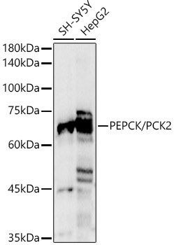
Western blot analysis of various lysates, using [KO Validated] PEPCK/PCK2 Rabbit pAb (CAB21529) at 1:1000 dilution. Secondary antibody: HRP-conjugated Goat anti-Rabbit IgG (H+L) (CABS014) at 1:10000 dilution. Lysates/proteins: 25μg per lane. Blocking buffer: 3% nonfat dry milk in TBST. Detection: ECL Basic Kit (AbGn00020). Exposure time: 3s.
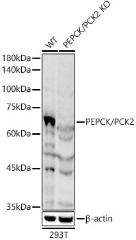
Western blot analysis of lysates from wild type(WT) and PEPCK/PCK2 Rabbit pAb knockout (KO) 293T cells, using [KO Validated] PEPCK/PCK2 Rabbit pAb (CAB21529) at 1:1000 dilution. Secondary antibody: HRP-conjugated Goat anti-Rabbit IgG (H+L) (CABS014) at 1:10000 dilution. Lysates/proteins: 25μg per lane. Blocking buffer: 3% nonfat dry milk in TBST. Detection: ECL Basic Kit (AbGn00020). Exposure time: 3s.
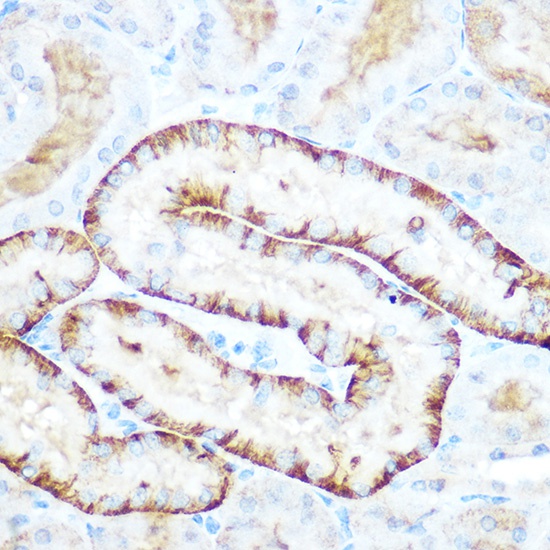
Immunohistochemistry analysis of paraffin-embedded Mouse kidney using [KO Validated] PEPCK/PCK2 Rabbit pAb (CAB21529) at dilution of 1:100 (40x lens). Microwave antigen retrieval performed with 0.01M Tris/EDTA Buffer (pH 9.0) prior to IHC staining.
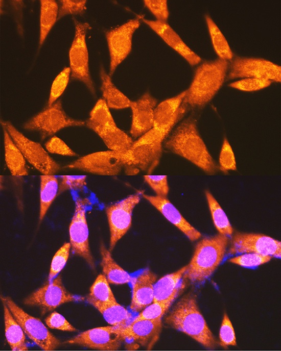
Immunofluorescence analysis of NIH-3T3 cells using PEPCK/PEPCK/PCK2 Rabbit pAb (CAB21529) at dilution of 1:100 (40x lens). Secondary antibody: Cy3-conjugated Goat anti-Rabbit IgG (H+L) (CABS007) at 1:500 dilution. Blue: DAPI for nuclear staining.
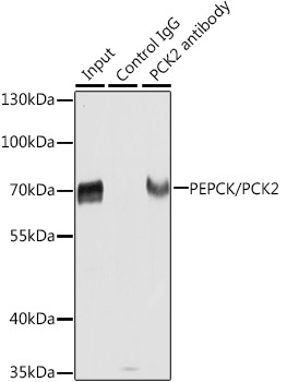
Immunoprecipitation analysis of extracts of HepG2 cells using PEPCK/PEPCK/PCK2 antibody (CAB21529). Western blot was performed from the immunoprecipitate using PEPCK/PEPCK/PCK2 antibody (CAB21529) at a dilution of 1:1000.
 at 1:1000 dilution. Secondary antibody: HRP Goat Anti-Rabbit IgG (H+L) at 1:10000 dilution. Lysates/proteins: 25μg per lane. Blocking buffer: 3% nonfat dry milk in TBST.")
 at 1:1000 dilution. Secondary antibody: HRP Goat Anti-Rabbit IgG (H+L) at 1:10000 dilution. Lysates/proteins: 25μg per lane. Blocking buffer: 3% nonfat dry milk in TBST.")


")

")
