The PFDN4 Antibody (CAB15300) is a high-quality antibody developed for reliable detection and analysis of target proteins. This antibody, generated in rabbits, exhibits high specificity and sensitivity for detecting PFDN4 in human samples, making it an essential tool for Western blot applications.PFDN4, also known as prefoldin subunit 4, plays a crucial role in maintaining protein homeostasis and preventing protein misfolding, which can lead to cellular dysfunction and disease. Its involvement in protein folding processes makes it a key player in various cellular functions, including cell growth, differentiation, and apoptosis.
This antibody is validated for use in WB, IF/ICC, ELISA applications and has demonstrated reactivity against Human, Mouse, Rat samples.
Product Name:
PFDN4 Antibody
SKU:
CAB15300
Size:
20μL, 100μL
Reactivity:
Human, Mouse, Rat
Conjugate:
Unconjugated
Immunogen:
Recombinant protein (or fragment).This information is considered to be commercially sensitive.
Sequence:
MAAT MKKA AAED VNVT FEDQ QKIN KFAR NTSR ITEL KEEI EVKK KQLQ NLED ACDD IMLA DDDC LMIP YQIG DVFI SHSQ EETQ EMLE EAKK NLQE EIDA LESR VESI QRVL ADLK VQLY AKFG SNIN LEAD ES
Tested Applications:
WBIF/ICCELISA
Recommended Dilution:
WB
1:200 - 1:2000
IF/ICC
1:50 - 1:200
ELISA
Recommended starting concentration is 1 μg/mL. Please optimize the concentration based on your specific assay requirements.
Synonyms:
C1, PFD4, PFDN4
Positive Sample:
DU145, HeLa, 293T, MCF7, mouse kidney
Cellular Localization:
Cytoplasm, Mitochondrion, Nucleus.
Calculated MW:
15kDa
Observed MW:
16kDa
This gene encodes a member of the prefoldin beta subunit family. The encoded protein is one of six subunits of prefoldin, a molecular chaperone complex that binds and stabilizes newly synthesized polypeptides, thereby allowing them to fold correctly. The complex, consisting of two alpha and four beta subunits, forms a double beta barrel assembly with six protruding coiled-coils.
Purification Method
Affinity purification
Gene ID
5203
RRID
AB_2762202
Buffer Information
Store at -20℃. Avoid freeze / thaw cycles. Buffer: PBS with 0.01% thimerosal,50% glycerol,pH7.3.
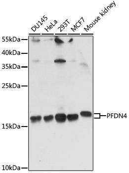
Western blot analysis of various lysates using PFDN4 Rabbit pAb (CAB15300) at 1:1000 dilution. Secondary antibody: HRP-conjugated Goat anti-Rabbit IgG (H+L) (CABS014) at 1:10000 dilution. Lysates/proteins: 25μg per lane. Blocking buffer: 3% nonfat dry milk in TBST. Detection: ECL Basic Kit (AbGn00020). Exposure time: 90s.
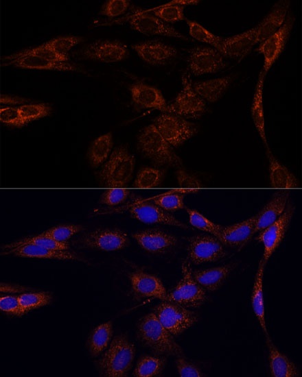
Immunofluorescence analysis of C6 cells using PFDN4 Rabbit pAb (CAB15300) at dilution of 1:100. Secondary antibody: Cy3-conjugated Goat anti-Rabbit IgG (H+L) (CABS007) at 1:500 dilution. Blue: DAPI for nuclear staining.
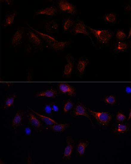
Immunofluorescence analysis of U-2 OS cells using PFDN4 Rabbit pAb (CAB15300) at dilution of 1:100. Secondary antibody: Cy3-conjugated Goat anti-Rabbit IgG (H+L) (CABS007) at 1:500 dilution. Blue: DAPI for nuclear staining.