The PFKL Antibody (CAB7708) is a high-quality antibody developed for reliable detection and analysis of target proteins. This antibody is produced in rabbits and has high reactivity with human samples, making it ideal for use in Western blot applications. By binding specifically to the PFKL protein, this antibody allows for accurate detection and analysis in a variety of cell types.PFKL is essential for ATP production and plays a crucial role in metabolic pathways. Its function in glycolysis makes it a target of interest in research on metabolic diseases, cancer metabolism, and energy metabolism in various tissues.
This antibody is validated for use in WB, IF/ICC, ELISA applications and has demonstrated reactivity against Human, Mouse, Rat samples.
Product Name:
PFKL Antibody
SKU:
CAB7708
Size:
20μL, 100μL
Reactivity:
Human, Mouse, Rat
Conjugate:
Unconjugated
Immunogen:
Recombinant protein (or fragment).This information is considered to be commercially sensitive.
Recommended starting concentration is 1 μg/mL. Please optimize the concentration based on your specific assay requirements.
Synonyms:
PFK-B, PFK-L, ATP-PFK, PFKL
Positive Sample:
Mouse heart, Rat heart
Cellular Localization:
Cytoplasm.
Calculated MW:
85kDa
Observed MW:
85kDa
This gene encodes the liver (L) subunit of an enzyme that catalyzes the conversion of D-fructose 6-phosphate to D-fructose 1,6-bisphosphate, which is a key step in glucose metabolism (glycolysis). This enzyme is a tetramer that may be composed of different subunits encoded by distinct genes in different tissues. Alternative splicing results in multiple transcript variants.
Purification Method
Affinity purification
Gene ID
5211
RRID
AB_2770868
Buffer Information
Store at -20℃. Avoid freeze / thaw cycles. Buffer: PBS containing 50% glycerol, preserved with proclin300 or sodium azide, pH 7.3.
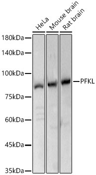
Western blot analysis of various lysates, using PFKL Rabbit pAb (CAB7708 ) at 1:3070 dilution. Secondary antibody: HRP-conjugated Goat anti-Rabbit IgG (H+L) (CABS014) at 1:10000 dilution. Lysates/proteins: 25μg per lane. Blocking buffer: 3% nonfat dry milk in TBST. Detection: ECL Basic Kit (AbGn00020). Exposure time: 1s.
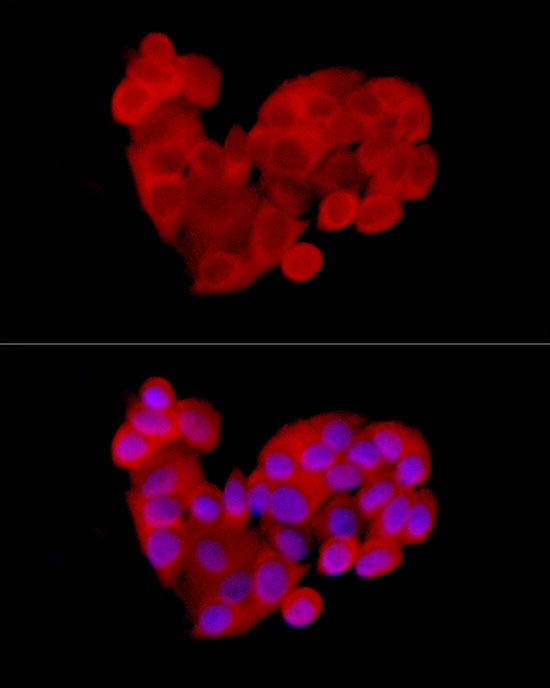
Immunofluorescence analysis of MCF7 cells using PFKL Rabbit pAb (CAB7708) at dilution of 1:300 (40x lens). Secondary antibody: Cy3-conjugated Goat anti-Rabbit IgG (H+L) (CABS007) at 1:500 dilution. Blue: DAPI for nuclear staining.
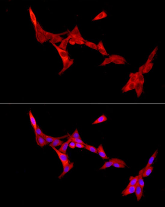
Immunofluorescence analysis of PC-12 cells using PFKL Rabbit pAb (CAB7708) at dilution of 1:300 (40x lens). Secondary antibody: Cy3-conjugated Goat anti-Rabbit IgG (H+L) (CABS007) at 1:500 dilution. Blue: DAPI for nuclear staining.