The PGD Antibody (CAB7710) is a high-quality antibody developed for reliable detection and analysis of target proteins. This antibody, produced in rabbits, is highly specific to human samples and has been validated for use in Western blot applications. By binding to PGD protein, this antibody enables accurate detection and analysis in a variety of cell types, making it an ideal choice for studies in biochemistry and inflammation research.
This antibody is validated for use in WB, IHC-P, IF/ICC, ELISA applications and has demonstrated reactivity against Human, Mouse, Rat samples.
Product Name:
PGD Antibody
SKU:
CAB7710
Size:
20μL, 100μL
Reactivity:
Human, Mouse, Rat
Conjugate:
Unconjugated
Immunogen:
Recombinant protein (or fragment).This information is considered to be commercially sensitive.
6-phosphogluconate dehydrogenase is the second dehydrogenase in the pentose phosphate shunt. Deficiency of this enzyme is generally asymptomatic, and the inheritance of this disorder is autosomal dominant. Hemolysis results from combined deficiency of 6-phosphogluconate dehydrogenase and 6-phosphogluconolactonase suggesting a synergism of the two enzymopathies. Several transcript variants encoding different isoforms have been found for this gene.
Purification Method
Affinity purification
Gene ID
5226
RRID
AB_2770874
Buffer Information
Store at -20℃. Avoid freeze / thaw cycles. Buffer: PBS containing 50% glycerol, preserved with proclin300 or sodium azide, pH 7.3.
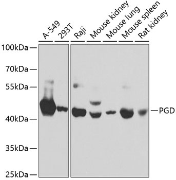
Western blot analysis of various lysates using PGD Rabbit pAb (CAB7710) at 1:1000 dilution. Secondary antibody: HRP-conjugated Goat anti-Rabbit IgG (H+L) (CABS014) at 1:10000 dilution. Lysates/proteins: 25μg per lane. Blocking buffer: 3% nonfat dry milk in TBST.
Immunohistochemistry analysis of paraffin-embedded Rat kidney using PGD Rabbit pAb (CAB7710) at dilution of 1:100 (40x lens). Microwave antigen retrieval performed with 0.01M PBS Buffer (pH 7.2) prior to IHC staining.
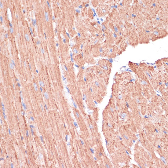
Immunohistochemistry analysis of paraffin-embedded Mouse heart using PGD Rabbit pAb (CAB7710) at dilution of 1:100 (40x lens). Microwave antigen retrieval performed with 0.01M PBS Buffer (pH 7.2) prior to IHC staining.
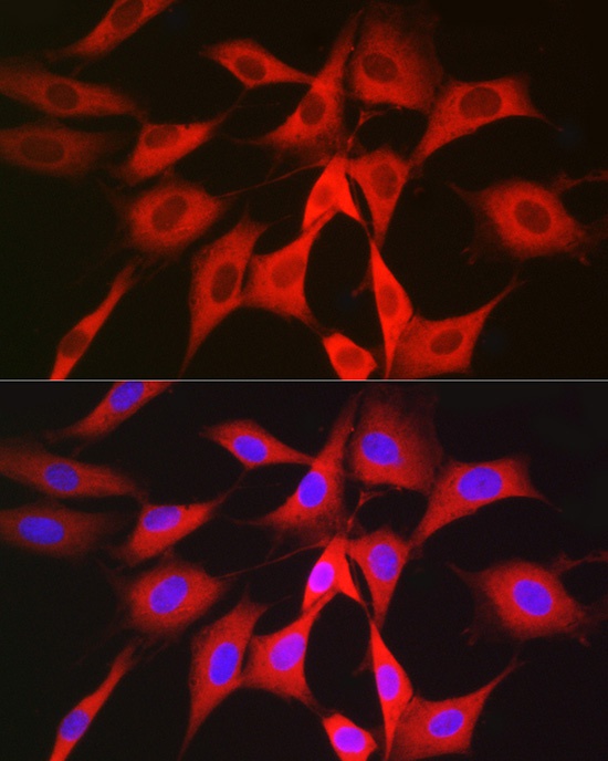
Immunofluorescence analysis of NIH/3T3 cells using PGD Rabbit pAb (CAB7710) at dilution of 1:50 (40x lens). Secondary antibody: Cy3-conjugated Goat anti-Rabbit IgG (H+L) (CABS007) at 1:500 dilution. Blue: DAPI for nuclear staining.
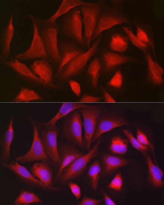
Immunofluorescence analysis of U2OS cells using PGD Rabbit pAb (CAB7710) at dilution of 1:50 (40x lens). Secondary antibody: Cy3-conjugated Goat anti-Rabbit IgG (H+L) (CABS007) at 1:500 dilution. Blue: DAPI for nuclear staining.