The Phospho-ATR-S428 Monoclonal Antibody (CABP1358) is a high-quality antibody developed for reliable detection and analysis of target proteins. ATR is a key regulator of the DNA damage response pathway, playing a critical role in maintaining genomic stability and preventing the accumulation of mutations that can lead to cancer. This monoclonal antibody, produced using cutting-edge technology, offers high sensitivity and specificity in detecting phosphorylated ATR at serine 428 in human samples. It is validated for use in Western blot applications, allowing for precise and reliable quantification of phospho-ATR levels in various cell types and experimental conditions.
This antibody is validated for use in WB, ELISA applications and has demonstrated reactivity against Human samples.
Product Name:
Phospho-ATR-S428 Monoclonal Antibody
SKU:
CABP1358
Size:
20μL, 100μL
Reactivity:
Human
Clone Number:
ARC55118
Conjugate:
Unconjugated
Immunogen:
Synthetic peptide. This information is considered to be commercially sensitive.
Sequence:
GISP K
Tested Applications:
WBELISA
Recommended Dilution:
WB
1:500 - 1:2000
ELISA
Recommended starting concentration is 1 μg/mL. Please optimize the concentration based on your specific assay requirements.
Synonyms:
FRP1, MEC1, SCKL, FCTCS, SCKL1, Phospho-ATR-S428
Positive Sample:
293T treated with UV
Cellular Localization:
Chromosome, Nucleus, Pml Body.
Calculated MW:
301kDa
Observed MW:
301kDa
The protein encoded by this gene is a serine/threonine kinase and DNA damage sensor, activating cell cycle checkpoint signaling upon DNA stress. The encoded protein can phosphorylate and activate several proteins involved in the inhibition of DNA replication and mitosis, and can promote DNA repair, recombination, and apoptosis. This protein is also important for fragile site stability and centrosome duplication. Defects in this gene are a cause of Seckel syndrome 1.
Purification Method
Affinity purification
Gene ID
545
Buffer Information
Store at -20℃. Avoid freeze / thaw cycles. Buffer: PBS containing 50% glycerol and 0.05% BSA, preserved with proclin300 or sodium azide, pH 7.3.
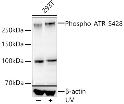
Western blot analysis of lysates from 293T cells using Phospho-ATR-S428 Rabbit mAb (CABP1358) at1:2000 dilution. 293T cells were treated with UV at room temperature for 15-30 minutes. Secondary antibody: HRP-conjugated Goat anti-Rabbit IgG (H+L) (CABS014) at 1:10000 dilution. Lysates/proteins: 30 μg per lane. Blocking buffer: 3% nonfat dry milk in TBST. Detection: ECL Basic Kit (AbGn00020). Exposure time: 90 s.
 at1:2000 dilution. 293T cells were treated by UV at room temperature for 15-30 minutes. Secondary antibody: HRP Goat Anti-Rabbit IgG (H+L) at 1:10000 dilution. Lysates/proteins: 25μg per lane. Blocking buffer: 3% nonfat dry milk in TBST.")
 at1:2000 dilution. 293T cells were treated by UV at room temperature for 15-30 minutes. Secondary antibody: HRP Goat Anti-Rabbit IgG (H+L) at 1:10000 dilution. Lysates/proteins: 25μg per lane. Blocking buffer: 3% nonfat dry milk in TBST.")


