The Phospho-c-Jun-S63 Monoclonal Antibody (CABP0105) is a high-quality antibody developed for reliable detection and analysis of target proteins. This antibody, produced in rabbits, is highly specific for detecting phosphorylated c-Jun at serine 63 in human samples, making it a reliable choice for Western blotting applications.Phosphorylation of c-Jun at serine 63 is known to regulate its activity and function in various cellular processes, including response to stress stimuli and activation of gene expression.
This antibody is validated for use in WB, IF/ICC, ELISA applications and has demonstrated reactivity against Human, Mouse, Rat samples.
Product Name:
Phospho-c-Jun-S63 Monoclonal Antibody
SKU:
CABP0105
Size:
20μL, 100μL
Reactivity:
Human, Mouse, Rat
Clone Number:
ARC0051
Conjugate:
Unconjugated
Immunogen:
Synthetic peptide. This information is considered to be commercially sensitive.
Sequence:
LTSP DV
Tested Applications:
WBIF/ICCELISA
Recommended Dilution:
WB
1:1000 - 1:10000
IF/ICC
1:50 - 1:200
ELISA
Recommended starting concentration is 1 μg/mL. Please optimize the concentration based on your specific assay requirements.
Synonyms:
AP1, p39, AP-1, cJUN, c-Jun, Phospho-c-Jun-S63
Positive Sample:
NIH/3T3 treated with Anisomycin, 293T treated with UV
Cellular Localization:
Nucleus.
Calculated MW:
36kDa
Observed MW:
48kDa
This gene is the putative transforming gene of avian sarcoma virus 17. It encodes a protein which is highly similar to the viral protein, and which interacts directly with specific target DNA sequences to regulate gene expression. This gene is intronless and is mapped to 1p32-p31, a chromosomal region involved in both translocations and deletions in human malignancies.
Purification Method
Affinity purification
Gene ID
3725
RRID
AB_2863804
Buffer Information
Store at -20℃. Avoid freeze / thaw cycles. Buffer: PBS containing 50% glycerol and 0.05% BSA, preserved with proclin300 or sodium azide, pH 7.3.
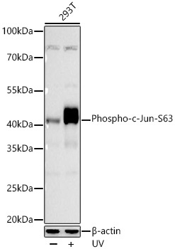
Western blot analysis of lysates from 293T cells using Phospho-c-Jun-S63 Rabbit mAb (CABP0105) at 1:10000 dilution incubated overnight at 4℃. 293T cells were treated with UV (100 mJ/cm2) at room temperature and recovered for 2 hours. Secondary antibody: HRP-conjugated Goat anti-Rabbit IgG (H+L) (CABS014) at 1:10000 dilution. Lysates/proteins: 30 μg per lane. Blocking buffer: 3% nonfat dry milk in TBST. Detection: ECL Basic Kit (AbGn00020). Exposure time: 45 s.
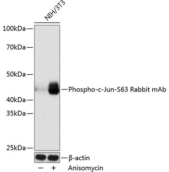
Western blot analysis of lysates from NIH/3T3 cells, using Phospho-c-Jun-S63 Rabbit mAb (CABP0105) at 1:1000 dilution. NIH/3T3 cells were treated with Anisomycin (25 μg/mL) at 37℃ for 30 minutes. Secondary antibody: HRP-conjugated Goat anti-Rabbit IgG (H+L) (CABS014) at 1:10000 dilution. Lysates/proteins: 25μg per lane. Blocking buffer: 3% nonfat dry milk in TBST. Detection: ECL Basic Kit (AbGn00020). Exposure time: 1s.