The Phospho-CDK4-T172 Antibody (CABP0593) is a high-quality antibody developed for reliable detection and analysis of target proteins. This rabbit-derived antibody specifically recognizes phosphorylated CDK4 at threonine 172, a key site involved in the activation of CDK4 and subsequent cell cycle progression.Validated for use in Western blot applications, this antibody is highly reactive with human samples, enabling precise detection and analysis of phospho-CDK4 levels in various cell types. Its specificity for phosphorylated CDK4 makes it an ideal tool for investigating the role of this protein kinase in cancer development and progression.
This antibody is validated for use in WB, ELISA, IF-P applications and has demonstrated reactivity against Human, Mouse, Rat samples.
Product Name:
Phospho-CDK4-T172 Antibody
SKU:
CABP0593
Size:
20μL, 100μL
Reactivity:
Human, Mouse, Rat
Conjugate:
Unconjugated
Immunogen:
Synthetic peptide. This information is considered to be commercially sensitive.
Sequence:
ALTP VV
Tested Applications:
WBELISAIF-P
Recommended Dilution:
WB
1:500 - 1:2000
IF-P
1:100 - 1:200
ELISA
Recommended starting concentration is 1 μg/mL. Please optimize the concentration based on your specific assay requirements.
Synonyms:
CMM3, PSK-J3, Phospho-CDK4-T172
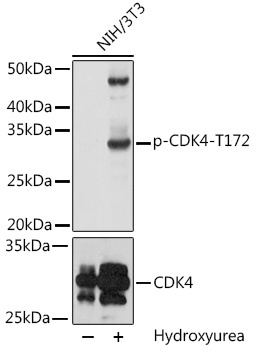
Positive Sample:
NIH/3T3 treated with Hydroxyurea
Cellular Localization:
Cytoplasm, Membrane, Nucleus.
Calculated MW:
34kDa
Observed MW:
33kDa
The protein encoded by this gene is a member of the Ser/Thr protein kinase family. This protein is highly similar to the gene products of S. cerevisiae cdc28 and S. pombe cdc2. It is a catalytic subunit of the protein kinase complex that is important for cell cycle G1 phase progression. The activity of this kinase is restricted to the G1-S phase, which is controlled by the regulatory subunits D-type cyclins and CDK inhibitor p16(INK4a). This kinase was shown to be responsible for the phosphorylation of retinoblastoma gene product (Rb). Mutations in this gene as well as in its related proteins including D-type cyclins, p16(INK4a) and Rb were all found to be associated with tumorigenesis of a variety of cancers. Multiple polyadenylation sites of this gene have been reported.
Purification Method
Affinity purification
Gene ID
1019
RRID
AB_2770981
Buffer Information
Store at -20℃. Avoid freeze / thaw cycles. Buffer: PBS with 0.09% Sodium azide,50% glycerol,pH7.3.
Western blot analysis of lysates from NIH/3T3 cells, using Phospho-CDK4-T172 Rabbit pAb (CAB0366). NIH/3T3 cells were treated with Hydroxyurea (4 mM) at 37℃ for 20 hours. Secondary antibody: HRP-conjugated Goat anti-Rabbit IgG (H+L) (CABS014) at 1:10000 dilution. Lysates/proteins: 25μg per lane. Blocking buffer: 3% BSA. Detection: ECL Basic Kit (AbGn00020). Exposure time: 60s.
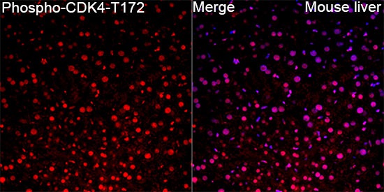
Immunofluorescence analysis of Mouse liver tissue using Phospho-CDK4-T172 Rabbit pAb (CABP0593) at a dilution of 1:100 (40x lens). Secondary antibody: Cy3-conjugated Goat anti-Rabbit IgG (H+L)(CABS007) at 1:500 dilution. Blue: DAPI for nuclear staining. High pressure antigen retrieval performed with 0.01M Citrate Buffer (pH 6.0) prior to IF staining.


")