The Phospho-EGFR-Y1172 Antibody (CABP0218) is a high-quality antibody developed for reliable detection and analysis of target proteins. This post-translational modification of EGFR is known to play a key role in signaling pathways involved in cell proliferation, survival, and migration.Our antibody is produced in rabbits and exhibits high reactivity with human samples, making it an ideal choice for Western blot applications. By specifically targeting the phosphorylated form of EGFR at Y1172, this antibody enables researchers to detect and analyze the activation status of EGFR in various cell types.
This antibody is validated for use in WB, ELISA applications and has demonstrated reactivity against Human samples.
Product Name:
Phospho-EGFR-Y1172 Antibody
SKU:
CABP0218
Size:
20μL, 100μL
Reactivity:
Human
Conjugate:
Unconjugated
Immunogen:
Synthetic peptide. This information is considered to be commercially sensitive.
Tested Applications:
WBELISA
Recommended Dilution:
WB
1:100 - 1:500
ELISA
Recommended starting concentration is 1 μg/mL. Please optimize the concentration based on your specific assay requirements.
Cell Membrane, Endoplasmic Reticulum Membrane, Endosome, Endosome Membrane, Golgi Apparatus Membrane, Nucleus Membrane, Nucleus, Secreted, Single-Pass Type I Membrane Protein.
Calculated MW:
134kDa
Observed MW:
175kDa
The protein encoded by this gene is a transmembrane glycoprotein that is a member of the protein kinase superfamily. This protein is a receptor for members of the epidermal growth factor family. EGFR is a cell surface protein that binds to epidermal growth factor, thus inducing receptor dimerization and tyrosine autophosphorylation leading to cell proliferation. Mutations in this gene are associated with lung cancer. EGFR is a component of the cytokine storm which contributes to a severe form of Coronavirus Disease 2019 (COVID-19) resulting from infection with severe acute respiratory syndrome coronavirus-2 (SARS-CoV-2).
Purification Method
Affinity purification
Gene ID
1956
RRID
AB_2771053
Buffer Information
Store at -20℃. Avoid freeze / thaw cycles. Buffer: PBS containing 50% glycerol, preserved with proclin300 or sodium azide, pH 7.3.
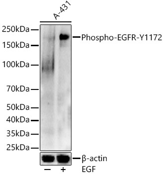
Western blot analysis of lysates from A-431 cells, using Phospho-EGFR-Y1172 Rabbit pAb (CABP0218) at 1:500 dilution. A-431 cells were treated with EGF (100 ng/ml) at 37℃ for 30 minutes after serum-starvation overnight. Secondary antibody: HRP-conjugated Goat anti-Rabbit IgG (H+L) (CABS014) at 1:10000 dilution. Lysates/proteins: 25μg per lane. Blocking buffer: 3% nonfat dry milk in TBST. Detection: ECL Enhanced Kit (AbGn00021). Exposure time: 60s.