The Phospho-JNK1/2/3-T183/T183/T221 Monoclonal Antibody (CABP0631) is a high-quality antibody developed for reliable detection and analysis of target proteins. This antibody, generated using a unique monoclonal antibody technology, specifically detects the phosphorylation of JNK1/2/3 at threonine residues 183/183/221 in human samples.The phosphorylation of JNK1/2/3 at these specific sites is known to be crucial for the activation of the JNK signaling pathway, which in turn regulates gene expression and cell survival in response to various stimuli.
This antibody is validated for use in WB, IHC-P, IF/ICC, ELISA applications and has demonstrated reactivity against Human, Mouse, Rat samples.
The protein encoded by this gene is a member of the MAP kinase family. MAP kinases act as an integration point for multiple biochemical signals, and are involved in a wide variety of cellular processes such as proliferation, differentiation, transcription regulation and development. This kinase is activated by various cell stimuli, and targets specific transcription factors, and thus mediates immediate-early gene expression in response to cell stimuli. The activation of this kinase by tumor-necrosis factor alpha (TNF-alpha) is found to be required for TNF-alpha induced apoptosis. This kinase is also involved in UV radiation induced apoptosis, which is thought to be related to cytochrom c-mediated cell death pathway. Studies of the mouse counterpart of this gene suggested that this kinase play a key role in T cell proliferation, apoptosis and differentiation. Several alternatively spliced transcript variants encoding distinct isoforms have been reported. [provided by RefSeq, Apr 2016]
Purification Method
Affinity purification
Gene ID
5599 5601 5602
RRID
AB_2771232
Buffer Information
Store at -20℃. Avoid freeze / thaw cycles. Buffer: PBS containing 50% glycerol and 0.05% BSA, preserved with proclin300 or sodium azide, pH 7.3.
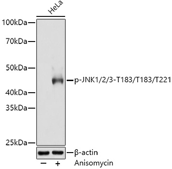
Western blot analysis of lysates from HeLa cells, using Phospho-JNK1/2/3-T183/T183/T221 Rabbit mAb (CABP0631) at 1:3000 dilution. HeLa cells were treated with Anisomycin (25 μg/mL) at 37℃ for 30 minutes after serum-starvation overnight. Secondary antibody: HRP-conjugated Goat anti-Rabbit IgG (H+L) (CABS014) at 1:10000 dilution. Lysates/proteins: 25μg per lane. Blocking buffer: 3% nonfat dry milk in TBST. Detection: ECL Basic Kit (AbGn00020). Exposure time: 180s.
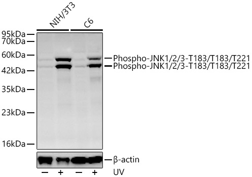
Western blot analysis of various lysates using Phospho-JNK1/2/3-T183/T183/T221 Rabbit mAb (CABP0631) at 1:1000 dilution incubated overnight at 4℃. NIH/3T3 cells and C6 cells were treated with UV at room temperature for 15-30 minutes. Secondary antibody: HRP-conjugated Goat anti-Rabbit IgG (H+L) (CABS014) at 1:10000 dilution. Lysates/proteins: 30 μg per lane. Blocking buffer: 3% nonfat dry milk in TBST. Detection: ECL Basic Kit (AbGn00020). Exposure time: 90s.
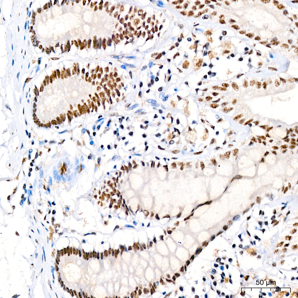
Immunohistochemistry analysis of paraffin-embedded Human colon using Phospho-JNK1/2/3-T183/T183/T221 Rabbit mAb (CABP0631) at dilution of 1:200 (40x lens). High pressure antigen retrieval performed with 0.01M Citrate buffer (pH 6.0) prior to IHC staining.
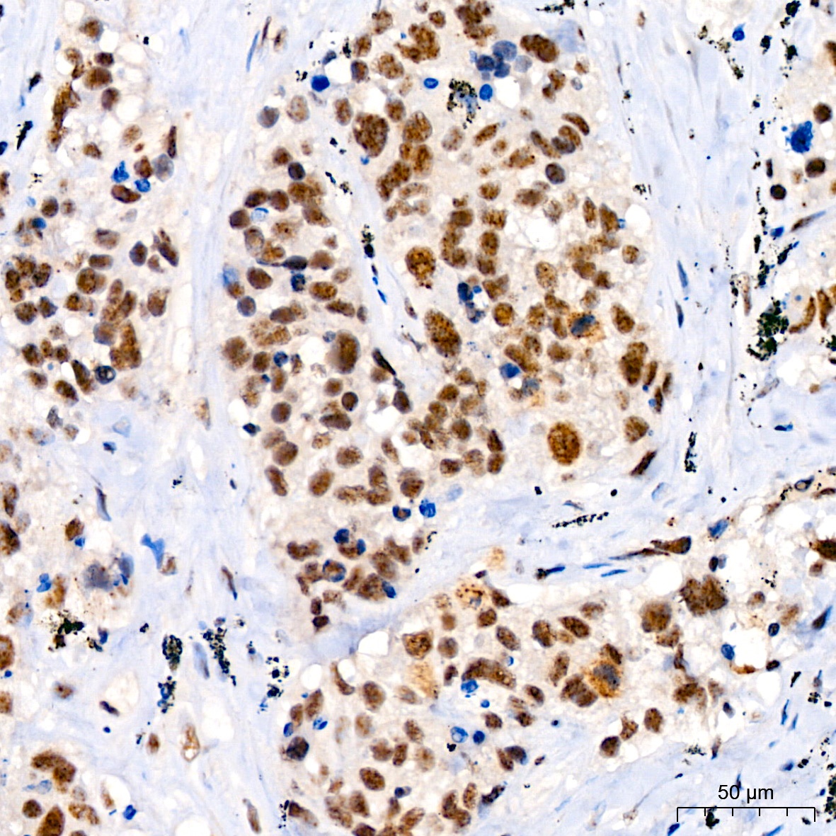
Immunohistochemistry analysis of paraffin-embedded Human lung squamous carcinoma tissue using Phospho-JNK1/2/3-T183/T183/T221 Rabbit mAb (CABP0631) at dilution of 1:200 (40x lens). High pressure antigen retrieval performed with 0.01M Citrate buffer (pH 6.0) prior to IHC staining.
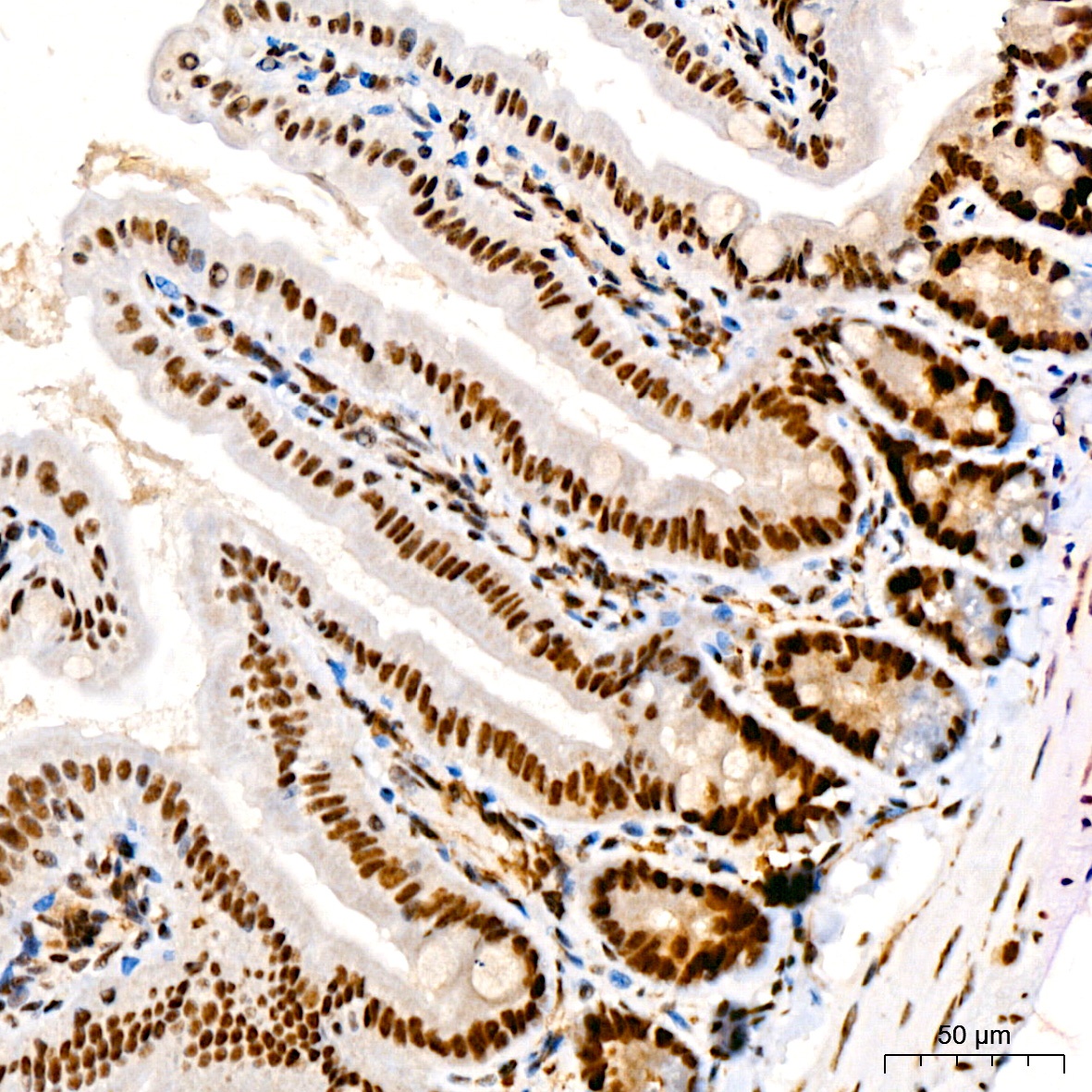
Immunohistochemistry analysis of paraffin-embedded Mouse colon using Phospho-JNK1/2/3-T183/T183/T221 Rabbit mAb (CABP0631) at dilution of 1:200 (40x lens). High pressure antigen retrieval performed with 0.01M Citrate buffer (pH 6.0) prior to IHC staining.
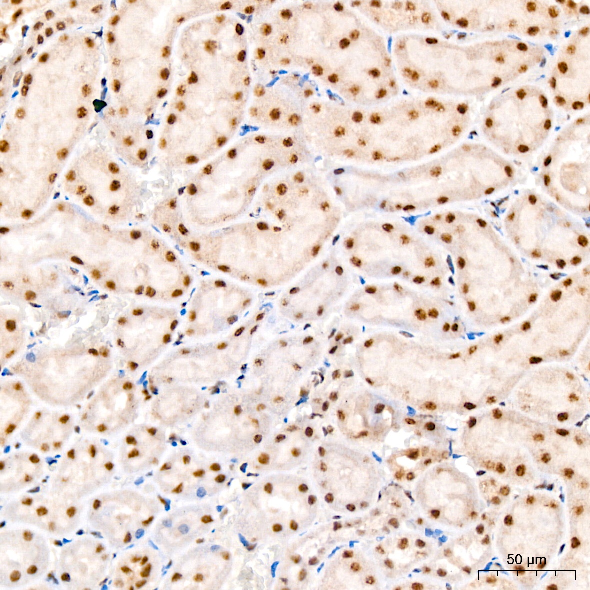
Immunohistochemistry analysis of paraffin-embedded Rat kidney using Phospho-JNK1/2/3-T183/T183/T221 Rabbit mAb (CABP0631) at dilution of 1:200 (40x lens). High pressure antigen retrieval performed with 0.01M Citrate buffer (pH 6.0) prior to IHC staining.
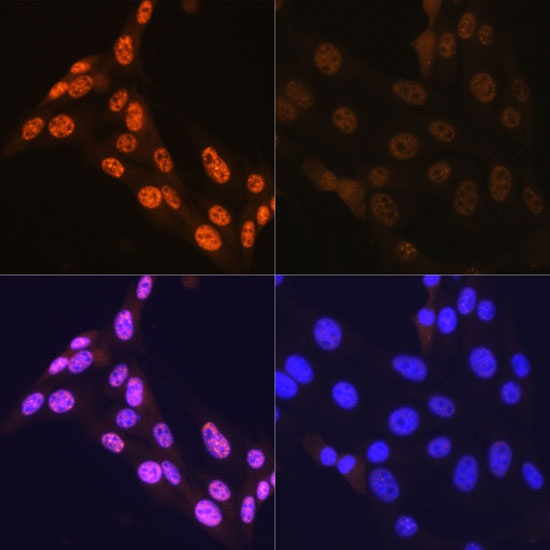
Immunofluorescence analysis of NIH-3T3 cells using Phospho-JNK1/2/3-T183/T183/T221 Rabbit mAb (CABP0631).NIH-3T3 cells were treated with Anisomycin (25 μg/mL) at 37℃ for 30 minutes after serum-starvation overnight. Secondary antibody: Cy3-conjugated Goat anti-Rabbit IgG (H+L) (CABS007) at 1:500 dilution. Blue: DAPI for nuclear staining.


")
