The Phospho-JunD-S255 Monoclonal Antibody (CABP1323) is a high-quality antibody developed for reliable detection and analysis of target proteins. JUN D is a transcription factor that plays a key role in regulating gene expression in response to various stimuli, including stress, cytokines, and growth factors. Phosphorylation at serine 255 is known to impact the activity and stability of JUN D, making it an important post-translational modification to study.This monoclonal antibody, produced using advanced technology, is highly specific for phosphorylated JUN D at serine 255 in human samples. It has been rigorously validated for use in Western blot applications, enabling precise detection and quantification of phosphorylated JUN D in various cell types and tissues.
This antibody is validated for use in WB, IHC-P, ELISA applications and has demonstrated reactivity against Human, Rat samples.
Product Name:
Phospho-JunD-S255 Monoclonal Antibody
SKU:
CABP1323
Size:
20μL, 100μL
Reactivity:
Human, Rat
Clone Number:
ARC54895
Conjugate:
Unconjugated
Immunogen:
Synthetic peptide. This information is considered to be commercially sensitive.
Sequence:
GESP P
Tested Applications:
WBIHC-PELISA
Recommended Dilution:
WB
1:500 - 1:2000
IHC-P
1:200 - 1:800
ELISA
Recommended starting concentration is 1 μg/mL. Please optimize the concentration based on your specific assay requirements.
Synonyms:
AP-1, Phospho-JunD-S255
Positive Sample:
HeLa
Cellular Localization:
Nucleus.
Calculated MW:
35kDa
Observed MW:
38kDa/42kDa
The protein encoded by this intronless gene is a member of the JUN family, and a functional component of the AP1 transcription factor complex. This protein has been proposed to protect cells from p53-dependent senescence and apoptosis. Alternative translation initiation site usage results in the production of different isoforms (PMID:12105216).
Purification Method
Affinity purification
Gene ID
3727
Buffer Information
Store at -20℃. Avoid freeze / thaw cycles. Buffer: PBS containing 50% glycerol and 0.05% BSA, preserved with proclin300 or sodium azide, pH 7.3.
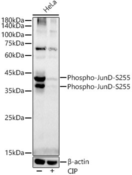
Western blot analysis of lysates from HeLa cells, using Phospho-JunD-S255 Rabbit mAb (CABP1323) at1:2000 dilution. Hela cells were treated by CIP(2 U/ul) at 37℃ for 1 hour. Secondary antibody: HRP-conjugated Goat anti-Rabbit IgG (H+L) (CABS014) at 1:10000 dilution. Lysates/proteins: 25 μg per lane. Blocking buffer: 3% nonfat dry milk in TBST. Detection: ECL Basic Kit (AbGn00020). Exposure time: 90 s.
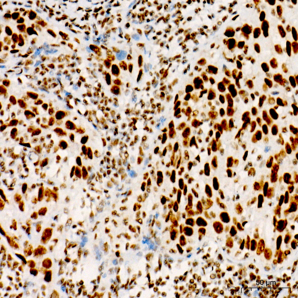
Immunohistochemistry analysis of paraffin-embedded Human cervix cancer tissue using Phospho-JunD-S255 Rabbit mAb (CABP1323) at a dilution of 1:500 (40x lens). High pressure antigen retrieval performed with 0.01M Citrate Buffer (pH 6.0) prior to IHC staining.
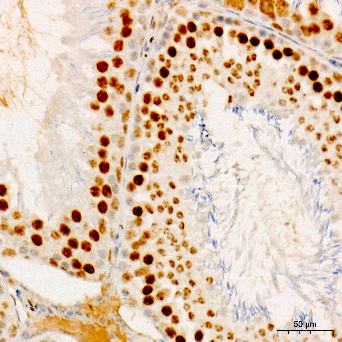
Immunohistochemistry analysis of paraffin-embedded Rat testis tissue using Phospho-JunD-S255 Rabbit mAb (CABP1323) at a dilution of 1:500 (40x lens). High pressure antigen retrieval performed with 0.01M Citrate Buffer (pH 6.0) prior to IHC staining.
 at1:2000 dilution. Hela cells were treated by CIP(20uL/400ul) at 37℃ for 1 hour. Secondary antibody: HRP Goat Anti-Rabbit IgG (H+L) at 1:10000 dilution. Lysates/proteins: 25μg per lane. Blocking buffer: 3% nonfat dry milk in TBST.")
 at1:2000 dilution. Hela cells were treated by CIP(20uL/400ul) at 37℃ for 1 hour. Secondary antibody: HRP Goat Anti-Rabbit IgG (H+L) at 1:10000 dilution. Lysates/proteins: 25μg per lane. Blocking buffer: 3% nonfat dry milk in TBST.")


