The Phospho-JunD-S255 Monoclonal Antibody (CABP1142) is a high-quality antibody developed for reliable detection and analysis of target proteins. JUND is a transcription factor that plays a crucial role in regulating gene expression in response to various stimuli, including stress, growth factors, and cytokines.This antibody specifically targets the phosphorylated form of JUND at serine 255, offering researchers a specific and sensitive tool for detecting and quantifying phosphorylated JUND in various cell types and experimental conditions. Its high reactivity with human samples, along with its validation for use in Western blot applications, make it an essential tool for studies in cell biology, signal transduction, and cancer research.
This antibody is validated for use in WB, ELISA applications and has demonstrated reactivity against Mouse samples.
Product Name:
Phospho-JunD-S255 Monoclonal Antibody
SKU:
CABP1142
Size:
20μL, 100μL
Reactivity:
Mouse
Clone Number:
ARC1603
Conjugate:
Unconjugated
Immunogen:
Synthetic peptide. This information is considered to be commercially sensitive.
Sequence:
GESP P
Tested Applications:
WBELISA
Recommended Dilution:
WB
1:500 - 1:2000
ELISA
Recommended starting concentration is 1 μg/mL. Please optimize the concentration based on your specific assay requirements.
Synonyms:
AP-1, Phospho-JunD-S255
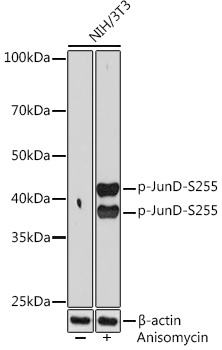
Positive Sample:
NIH/3T3 treated with Anisomycin
Cellular Localization:
Nucleus.
Calculated MW:
35kDa
Observed MW:
38kDa/42kDa
The protein encoded by this intronless gene is a member of the JUN family, and a functional component of the AP1 transcription factor complex. This protein has been proposed to protect cells from p53-dependent senescence and apoptosis. Alternative translation initiation site usage results in the production of different isoforms (PMID:12105216).
Purification Method
Affinity purification
Gene ID
3727
RRID
AB_2864009
Buffer Information
Store at -20℃. Avoid freeze / thaw cycles. Buffer: PBS containing 50% glycerol and 0.05% BSA, preserved with proclin300 or sodium azide, pH 7.3.
Western blot analysis of lysates from NIH/3T3 cells, using Phospho-JunD-S255 Rabbit mAb (CABP1142) at 1:1000 dilution. NIH/3T3 cells were treated with Anisomycin (25 μg/mL) at 37℃ for 30 minutes. Secondary antibody: HRP-conjugated Goat anti-Rabbit IgG (H+L) (CABS014) at 1:10000 dilution. Lysates/proteins: 25μg per lane. Blocking buffer: 3% BSA. Detection: ECL Basic Kit (AbGn00020). Exposure time: 90s.