The Phospho-c-Myc-T58 Antibody (CABP0080) is a high-quality antibody developed for reliable detection and analysis of target proteins. This post-translational modification plays a key role in regulating Myc protein stability, transcriptional activity, and ultimately cell proliferation.Raised in rabbits, this antibody is highly specific for human samples and has been validated for use in Western blot applications, allowing for precise detection and quantification of phospho-Myc (T58) levels in various cell types. Its high reactivity and specificity make it an ideal choice for studies focused on understanding the signaling pathways involving Myc protein and its role in cancer progression.The phosphorylation of Myc at threonine 58 has been linked to the dysregulation of cell growth and survival in numerous cancer types, making it a promising target for therapeutic intervention.
This antibody is validated for use in WB, IHC-P, ELISA applications and has demonstrated reactivity against Human, Mouse, Rat samples.
Product Name:
Phospho-c-Myc-T58 Antibody
SKU:
CABP0080
Size:
20μL, 100μL
Reactivity:
Human, Mouse, Rat
Conjugate:
Unconjugated
Immunogen:
Synthetic peptide. This information is considered to be commercially sensitive.
Sequence:
LPTP P
Tested Applications:
WBIHC-PELISA
Recommended Dilution:
WB
1:100 - 1:500
IHC-P
1:50 - 1:200
ELISA
Recommended starting concentration is 1 μg/mL. Please optimize the concentration based on your specific assay requirements.
Synonyms:
MRTL, MYCC, c-Myc, bHLHe39, Phospho-c-Myc-T58
Positive Sample:
293T treated with Calyculin A
Cellular Localization:
Nucleus, Nucleolus, Nucleoplasm.
Calculated MW:
51kDa
Observed MW:
62kDa
This gene is a proto-oncogene and encodes a nuclear phosphoprotein that plays a role in cell cycle progression, apoptosis and cellular transformation. The encoded protein forms a heterodimer with the related transcription factor MAX. This complex binds to the E box DNA consensus sequence and regulates the transcription of specific target genes. Amplification of this gene is frequently observed in numerous human cancers. Translocations involving this gene are associated with Burkitt lymphoma and multiple myeloma in human patients. There is evidence to show that translation initiates both from an upstream, in-frame non-AUG (CUG) and a downstream AUG start site, resulting in the production of two isoforms with distinct N-termini.
Purification Method
Affinity purification
Gene ID
4609
RRID
AB_2771348
Buffer Information
Store at -20℃. Avoid freeze / thaw cycles. Buffer: PBS with 0.09% Sodium azide,50% glycerol,pH7.3.
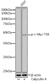
Western blot analysis of lysates from 293T cells, using Phospho-c-Myc-T58 Rabbit pAb (CABP0080) at 1:500 dilution. 293T cells were treated with Calyculin A (100 nM) at 37℃ for 30 minutes after serum-starvation overnight. Secondary antibody: HRP-conjugated Goat anti-Rabbit IgG (H+L) (CABS014) at 1:10000 dilution. Lysates/proteins: 25μg per lane. Blocking buffer: 3% nonfat dry milk in TBST. Detection: ECL Enhanced Kit (AbGn00021). Exposure time: 180s.
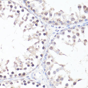
Immunohistochemistry analysis of paraffin-embedded Rat testis using Phospho-c-Myc-T58 Rabbit pAb (CABP0080) at dilution of 1:100 (40x lens). Microwave antigen retrieval performed with 0.01M Tris/EDTA Buffer (pH 9.0) prior to IHC staining.
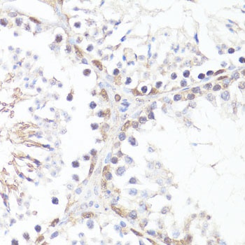
Immunohistochemistry analysis of paraffin-embedded Mouse testis using Phospho-c-Myc-T58 Rabbit pAb (CABP0080) at dilution of 1:100 (40x lens). Microwave antigen retrieval performed with 0.01M Tris/EDTA Buffer (pH 9.0) prior to IHC staining.